. 2025 Feb;21(2):e14433.
doi: 10.1002/alz.14433. Epub 2025 Jan 3.

Retinal dysfunction in APOE4 knock-in mouse model of Alzheimer's disease

Affiliations

Retinal dysfunction in APOE4 knock-in mouse model of Alzheimer's disease

Surabhi D Abhyankar et al. Alzheimers Dement. 2025 Feb.

Abstract

Introduction: Late-onset Alzheimer's Disease (LOAD) is the predominant form of Alzheimer's disease (AD), and apolipoprotein E (APOE) ε4 is a strong genetic risk factor for LOAD. As an integral part of the central nervous system, the retina displays a variety of abnormalities in LOAD. Our study is focused on age-dependent retinal impairments in humanized APOE4-knock-in (KI) and APOE3-KI mice developed by the Model Organism Development and Evaluation for Late-Onset Alzheimer's Disease (MODEL-AD) consortium.

Methods: All the experiments were performed on 52- to 57-week-old mice. The retina was assessed by optical coherence tomography, fundoscopy, fluorescein angiography, electroretinography, optomotor response, gliosis, and neuroinflammation. mRNA sequencing was performed to find molecular pathways.

Results: APOE4-KI mice showed impaired retinal structure, vasculature, function, vision, increased gliosis and neuroinflammation, and downregulation of synaptogenesis.

Discussion: The APOE ε4 allele is associated with increased susceptibility to retinal degeneration compared to the APOE ε3 allele.

Highlights: Apolipoprotein E (APOE)4 mice exhibit structural and functional deficits of the retina. The retinal defects in APOE4 mice are attributed to increased neuroinflammation. APOE4 mice show a unique retinal transcriptome, yet with key brain similarities. The retina offers a non-invasive biomarker for the detection and monitoring of Alzheimer's disease.

Keywords: APOE; Late‐Onset Alzheimer's Disease; biomarker; retina; retinal dysfunction.


Conflict of interest statement

A.B. is an ad hoc District Support Pharmacist at CVS Health/Aetna. The contents of this study do not reflect those of CVS Health/Aetna. Q.L., G.H., N.M., T.W.C., A.L.O., B.T.L., and S.A. do not have any conflicts to declare. Author disclosures are available in the Supporting information.

Figures
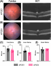
FIGURE 1
FIGURE 1
Reduced retinal thickness in APOE4 mice. (A, B) Representative fundus and OCT images of APOE3 and APOE4, respectively, Scale bars: 100 µm. (C) Total retina thickness. (D) Inner retina thickness. (E) Outer retina thickness. Values are expressed as mean ± SEM. (n: APOE3 = 14, APOE4 = 11). Unpaired t‐test. *p < 0.05, ****p < 0.0001.
FIGURE 2
FIGURE 2
Vascular tortuosity in APOE4 mice. (A, B) Representative fundus images of fluorescein angiography (FA) and vascular area of APOE3 and APOE4, respectively, and (C) tortuosity (indicated with white arrows). (D) Largest retinal vein width. (E) Vascular area. (F) Avascular area. Values are expressed as mean ± SEM. (n: APOE3 = 14, APOE4 = 11). Unpaired t‐test. *p < 0.05.
FIGURE 3
FIGURE 3
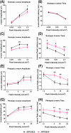
APOE4 mice have functional deficits in their retinas. a‐wave amplitude (A), a‐wave time (B), b‐wave amplitude (C), and b‐wave time (D) under scotopic conditions. a‐wave amplitude (E), a‐wave time (F), b‐wave amplitude (G), and b‐wave time (H) under photopic conditions. Values are expressed as mean ± SEM (n: APOE3 = 18, APOE4 = 21). Two‐way ANOVA with Fisher's multiple comparisons test. *p < 0.05, **p < 0.01, ***p < 0.001.
FIGURE 4
FIGURE 4
APOE4 mice have lower visual acuity (A) and contrast sensitivity (B). Values are expressed as mean ± SEM (n: APOE3 = 14, APOE4 = 12). Unpaired t‐test. *p < 0.05.
FIGURE 5
FIGURE 5
APOE4 mice have increased GFAP expression and increased inflammation. (A) Representative images of retinal slices showing GFAP staining pattern in APOE3 and APOE4 mice. (n: APOE3 = 3, APOE4 = 3. GCL, ganglion cell layer; IPL, inner plexiform layer; INL, inner nuclear layer; OPL, outer plexiform layer; ONL, outer nuclear layer. (B) Real‐time qRT‐PCR results showing mRNA expression of IL6 and IL1β normalized to the housekeeping gene TBP. (n: APOE3 = 8, APOE4 = 8). Values are expressed as mean ± SEM. Student's unpaired t‐test. *p < 0.05, **p < 0.01.
FIGURE 6
FIGURE 6
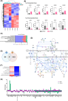
APOE4 retinas showed downregulation of synaptogenesis. (A) Heatmap showing significantly expressed genes in APOE4 retinas. (B) mRNA validation of top five upregulated and top five downregulated genes by qRT‐PCR normalized to the housekeeping gene TBP. (n: 6–8 mice/group). (C) Top 10 canonical pathways affected in APOE4 retinas. (D) Synaptogenesis signaling pathway. DEGs from RNA sequencing were superimposed on all the targets in the pathway. Red indicates upregulated genes, whereas green indicates downregulated genes. The degree of up‐ or down‐regulation is shown by the intensity of red and green colors, respectively. Predicted inhibition is indicated by blue. (E) Venn diagram representing a common number of genes between the brain and retina. (F) Heatmap showing top canonical pathways between brain and retina of APOE4. (G) Bar graph showing fold change of 97 common genes between brain and retina of APOE4.

Similar articles

Cited by

References

    1. Madnani RS. Alzheimer's disease: a mini‐review for the clinician. Front Neurol. 2023;14:1178588. - PMC - PubMed
    1. Li Y, Laws SM, Miles LA, et al. Genomics of Alzheimer's disease implicates the innate and adaptive immune systems. Cell Mol Life Sci. 2021;78(23):7397‐7426. - PMC - PubMed
    1. Yokoyama M, Kobayashi H, Tatsumi L, Tomita T. Mouse models of Alzheimer's disease. Front Mol Neurosci. 2022;15:912995. - PMC - PubMed
    1. Zhong MZ, Peng T, Duarte ML, Wang M, Cai D. Updates on mouse models of Alzheimer's disease. Mol. 2024;19(1):23. - PMC - PubMed
    1. Bales KR. The value and limitations of transgenic mouse models used in drug discovery for Alzheimer's disease: an update. Expert Opin Drug Discov. 2012;7(4):281‐297. - PubMed

Substances