. 2025 Apr;66(4):1234-1249.
doi: 10.1111/epi.18251. Epub 2025 Jan 3.

Detecting somatic variants in purified brain DNA obtained from surgically implanted depth electrodes in epilepsy

Collaborators, Affiliations

Detecting somatic variants in purified brain DNA obtained from surgically implanted depth electrodes in epilepsy

Rumika Mascarenhas et al. Epilepsia. 2025 Apr.

Abstract

Objective: Somatic variants causing epilepsy are challenging to detect, as they are only present in a subset of brain cells (e.g., mosaic), resulting in low variant allele frequencies. Traditional methods relying on surgically resected brain tissue are limited to patients undergoing brain surgery. We developed an improved protocol to detect somatic variants using DNA from stereoelectroencephalographic (SEEG) depth electrodes, enabling access to a larger patient cohort and diverse brain regions. This protocol mitigates issues of contamination and low yields by purifying neuronal nuclei using fluorescence-activated nuclei sorting (FANS).

Methods: SEEG depth electrodes were collected upon extraction from 41 brain regions across 17 patients undergoing SEEG. Nuclei were isolated separately from depth electrodes in the affected brain regions (seizure onset zone) and the unaffected brain regions. Neuronal nuclei were isolated using FANS, and DNA was amplified using primary template amplification. Short tandem repeat (STR) analysis and postsequencing allelic imbalance assessment were used to evaluate sample integrity. High-quality amplified DNA samples from affected brain regions, patient-matched unaffected brain regions, and genomic DNA were subjected to whole exome sequencing (WES). A bioinformatic workflow was developed to reduce false positives and to accurately detect somatic variants in the affected brain region.

Results: Based on DNA yield and STR analysis, 14 SEEG-derived neuronal DNA samples (seven affected and seven unaffected) across seven patients underwent WES. From the variants prioritized using our bioinformatic workflow, we chose four candidate variants in MTOR, CSDE1, KLLN, and NLE1 across four patients based on pathogenicity scores and association with phenotype. All four variants were validated using digital droplet polymerase chain reaction.

Significance: Our approach enhances the reliability and applicability of SEEG-derived DNA for epilepsy, offering insights into its molecular basis, facilitating epileptogenic zone identification, and advancing precision medicine.

Keywords: DNA; depth electrodes; epilepsy; somatic variant; stereo‐EEG.


Conflict of interest statement

The authors declare no conflicts of interest.

Figures
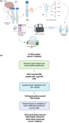
FIGURE 1
FIGURE 1
Overview of methodology for stereoelectroencephalography (SEEG)‐derived DNA analysis to detect somatic brain variants. (A) Workflow summarizing the steps involved in DNA extraction from neuronal nuclei isolated from SEEG depth electrodes to detect somatic brain variants. (B) Flowchart showing how SEEG samples were selected for whole exome sequencing. ddPCR, digital droplet polymerase chain reaction; FANS, fluorescence‐activated nuclei sorting; STR, short tandem repeat.
FIGURE 2
FIGURE 2
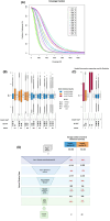
Postsequencing quality control (QC) measures and variant filtration workflow. (A) Coverage fraction. This panel illustrates which fraction of the exome (reference) was covered at which sequencing depth (coverage) for the sequenced stereoelectroencephalography (SEEG)‐derived neuronal DNA samples. *A, affected brain region, U, unaffected brain region, G, germline DNA from blood/saliva. (B, C) Analysis of allelic imbalance in SEEG‐derived neuronal DNA and germline‐derived DNA samples. These panels present the distribution of variant allele frequency (VAF) among heterozygous variants identified in SEEG‐derived neuronal DNA samples (affected and unaffected) compared with germline‐derived DNA samples (blood/saliva). The box plot displays the median (center line), interquartile range (box limits at the 25th and 75th percentiles), whiskers extending to 1.5 times the interquartile range, and individual points representing outliers. (B) Data from seven patients included in this publication. (C) Data from three patients from an earlier sequencing round, before optimizing our SEEG harvesting protocol, to demonstrate the allelic imbalance quality in samples with suboptimal short tandem repeat (STR) quality. A, affected SEEG‐derived neuronal DNA samples; G, germline‐derived DNA samples; U, unaffected SEEG‐derived neuronal DNA samples. The allelic imbalance quality is classified as follows: Level A for patient samples with affected and unaffected neuronal samples falling below the 1 × SD threshold of 11.5%, Level B for patient samples with unaffected and/or affected samples in between the 1 × and 2 × SD thresholds (11.5%–23.0%), and Level C for patient samples with unaffected and/or affected samples exceeding the 2 × SD threshold (23.0%). #The STR analysis is categorized as "perfect" (no allelic dropout), “good” (≤2 markers with dropout), “medium” (≤4 markers with dropout), and "low" (>4 markers with dropout). These visualizations were generated using the ggplot2 package in R. (D) Schematic illustrating the stepwise filtration process of variants from raw VCF files generated by Mutect2 and the corresponding average number of variants retained at each stage for Level A and Level B quality samples. Starting with raw variant calls, the process undergoes successive refinement through five major steps: (1) filtration using FilterMutectCalls; variants with the “PASS” flag were retained; (2) artifact removal; based on in‐house database, variants in <2 other neuronal samples were retained; (3) population database‐based filtration; variants with <1% population frequency in GnomADv2 were retained; (4) gene panel and pathogenicity‐based filtration: variants in epilepsy genes and/or variants with Combined Annotation Dependent Depletion score > 12 were retained; and (5) manual review using Integrative Genome Viewer (IGV; see Materials and Methods for details), culminating in the selection of prioritized variants. The average number of variants retained after each step is displayed (in black font), alongside the average percentage reduction observed during each filtration step (in red font).
FIGURE 3
FIGURE 3
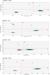
Digital droplet polymerase chain reaction (ddPCR) results for candidate variants in affected brain regions showing variant confirmation in affected samples. Due to technical issues (GC‐rich region), the KLLN assay separated less well between positive and negative variant droplets. Gating was determined based on fluorescent intensities of no template control samples. Blue: variant droplets, green: wild‐type droplets, orange: droplets containing multiple DNA templates, gray: empty droplets. Red arrows show bulk of variant droplets for CSDE1, NLE1, KLLN, and MTOR. ddPCR results for candidate variants in unaffected brain regions and germline samples are available as Figure S1.

References

    1. Lee JH, Huynh M, Silhavy JL, Kim S, Dixon‐Salazar T, Heiberg A, et al. De novo somatic mutations in components of the PI3K‐AKT3‐mTOR pathway cause hemimegalencephaly. Nat Genet. 2012;44:941–945. - PMC - PubMed
    1. Lim JS, Kim WI, Kang HC, Kim SH, Park AH, Park EK, et al. Brain somatic mutations in MTOR cause focal cortical dysplasia type II leading to intractable epilepsy. Nat Med. 2015;21:395–400. - PubMed
    1. Lim JS, Gopalappa R, Kim SH, Ramakrishna S, Lee M, Kim WI, et al. Somatic mutations in TSC1 and TSC2 cause focal cortical dysplasia. Am J Hum Genet. 2017;100:454–472. - PMC - PubMed
    1. Baldassari S, Ribierre T, Marsan E, Adle‐Biassette H, Ferrand‐Sorbets S, Bulteau C, et al. Dissecting the genetic basis of focal cortical dysplasia: a large cohort study. Acta Neuropathol (Berl). 2019;138:885–900. - PMC - PubMed
    1. D'Gama AM, Woodworth MB, Hossain AA, Bizzotto S, Hatem NE, LaCoursiere CM, et al. Somatic mutations activating the mTOR pathway in dorsal Telencephalic progenitors cause a continuum of cortical Dysplasias. Cell Rep. 2017;21:3754–3766. - PMC - PubMed