. 2025 Jan 3;74(2):66.
doi: 10.1007/s00262-024-03907-y.

Characterization of the adaptive immune response in a mouse model for HPV-positive head and neck squamous cell carcinoma with implications to human disease

Affiliations

Characterization of the adaptive immune response in a mouse model for HPV-positive head and neck squamous cell carcinoma with implications to human disease

Franziska Oliveri et al. Cancer Immunol Immunother. .

Abstract

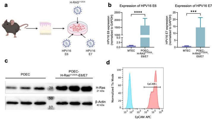
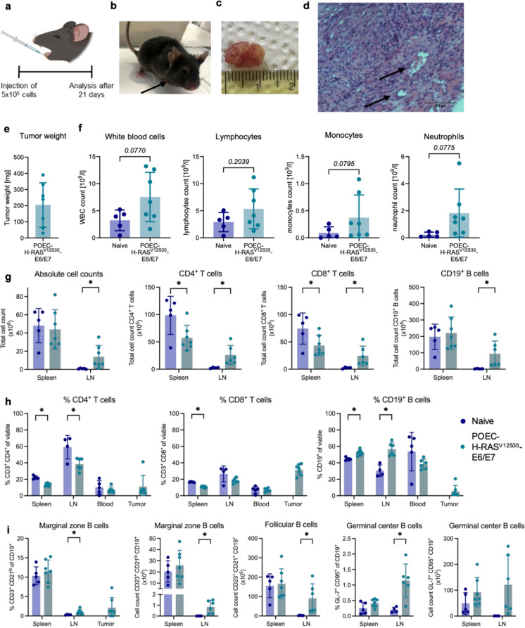
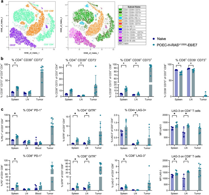
Head and neck squamous cell carcinoma (HNSCC) is the seventh most common cancer worldwide with a poor prognosis for survival. Risk factors include alcohol and tobacco abuse and infection with human papilloma virus (HPV). To enhance anti-tumor immune responses immunotherapeutic approaches are approved for recurrent metastatic disease but only approx. 20% of patients respond to checkpoint blockade of the PD-1/PD-L1 axis. Therefore, preclinical research is needed to better understand molecular and cellular processes and to identify new therapeutic targets. Immunocompetent mouse models can serve these purposes but only few are currently available for HPV-positive HNSCC. Here, we established a mouse cell line overexpressing the oncogenes E6/E7 of the HPV16 genome as well as a constitutively active form of H-Ras and studied the anti-tumor immune response upon orthotopic tumor growth at the floor of the mouth. Moreover, we analyzed the same immunoregulatory pathways in samples of HPV-positive cancer patients. T cells in the tumor of mice and humans exhibited high expression of CD39 and CD73, two ectoenzymes involved in the production of immunosuppressive adenosine from ATP, along with increased expression of PD-1, LAG-3 and GITR. Additionally, B cell responses were elevated in tumor-bearing mice, seen as an increase of germinal center, immunoregulatory marginal zone and follicular B cell subtypes. Taken together, this study suggests that the generated mouse model shares characteristics with human disease and can thus be used as a platform to study anti-tumor responses in HPV-positive HNSCC which will help to identify novel therapeutic targets.

Keywords: HPV; Head and neck cancer; Immune checkpoints; Tumor-infiltrating lymphocytes.


Conflict of interest statement

Declarations. Conflict of interest: The authors declare no competing interests. Ethics approval: This study was performed in line with the principles of the Declaration of Helsinki. Approval was granted by the Ethics Committee of Ulm University (Votum #90/15). Consent to participate: All donors of the present study provided informed and written consent. Animal experimentation was approved by the Regierungspräsidium Tübingen (permission number #1607).

Figures

Fig. 1
Fig. 1
Generation of a murine cell line to mimic HPV-positive HNSCC. Murine primary oral epithelial cells (POECs) were isolated from oral mucosal tissue of wildtype C56BL/6 mice and retrovirally transduced for the overexpression of H-RasV12S35 and E6/E7 of the human papilloma virus type 16 (HPV16) genome. a Schematic overview of the procedure (created by BioRender.com). b RNA expression levels of HPV16-E6 (left) and E7 (right) in original and modified POECs analyzed by qPCR and normalized to the house keeping gene GAPDH. c Protein levels of H-Ras in three samples of original and modified POECs analyzed by immunoblotting. Note that wildtype H-Ras was detected due to a lack of a mutation-specific antibody. β-actin served as loading control. d Flow cytometric analysis of EpCAM expression on POEC- H-RAS.V12S35-E6/E7. Blue histogram shows unstained control. Statistical significance of differences was determined using unpaired Student’s t tests and is depicted as p < 0.001 (***), p < 0.0001 (****)
Fig. 2
Fig. 2
Characterization of T and B cell responses in POEC- H-RASV12S35-E6/E7 tumor-bearing mice. 5 × 105 POEC- H-RASV12S35-E6/E7 cells were implanted into the floor of the mouth of C57BL/6 mice. Tumor growth and the immune response was analyzed after 21 days. a Experimental setup (created by BioRender.com). b Representative photograph of a tumor-bearing mouse on day 21 before analysis. c Representative pictures of dissected tumors and d micrographs of hematoxylin/eosin-stained tumor tissue. Arrows indicate blood vessels. e Tumor weight of dissected tumors. f Blood analysis using VetSan HM5 of naïve vs. tumor-bearing for the analysis of white blood cells (WBC), lymphocytes, monocytes and neutrophils. g Absolute cell counts of all cells and CD4+ and CD8+ T cells as well as CD19+ B cells in the spleen and lymph nodes in naïve compared to tumor-bearing mice determined by flow cytometry. h Relative proportions of T and B cells as well as i different B cells subsets in spleen, lymph node (LN), blood and tumor determined by flow cytometry. All cellular subsets were identified as specified on the y-axis of the graphs after gating on the lymphocyte population and exclusion of debris and dead cells. Data are shown as mean ± SD, and each point represents an individual mouse (n = 4–7). Statistical significance of differences between naïve and tumor-bearing mice was determined by unpaired Student’s t tests or Mann–Whitney U test for parametric or nonparametric data, respectively. Statistical significance is depicted as p < 0.05 (*) or given as full numbers of exact p-values
Fig. 3
Fig. 3
Immunoregulatory pathways are enhanced in POEC-H-RASV12S35-E6/E7 tumor-bearing mice. 5 × 105 POEC- H-RASV12S35-E6/E7 cells were implanted into the floor of the mouth of C57BL/6 mice, and the immune response was analyzed after 21 days. a Representative tSNE blots of viable immune cells in POEC- H-RASV12S35-E6/E7 tumors analyzed by spectral flow cytometry. Different immune cell populations were identified by traditional gating schemes and overlayed in different colors to visualize population distributions (legend on the right). b CD39 and/or CD73-expressing T cell subsets in spleen, lymph node (LN) and tumor. c CD4+ (upper panel) and CD8+ (lower panel) T cell subsets expressing various immunoregulatory receptors in the different organs. Data is shown as relative proportion of positive cells for all subsets and additionally as mean fluorescence intensity (MFI) for LAG-3 (right side). All cellular subsets were identified as specified on the y-axis of the graphs after gating on the lymphocyte population and exclusion of debris and dead cells. Data are shown as mean ± SD, and each point represents an individual mouse (n = 4–7). Statistical significance of differences between naïve and tumor-bearing mice was determined by unpaired student’s t tests or Mann–Whitney U test for parametric or nonparametric data, respectively, and depicted as p < 0.05 (*)
Fig. 4
Fig. 4
Immunoregulatory pathways in human HPV + HNSCC. Peripheral blood mononuclear cells (PBMCs) were obtained from healthy donors, and patients with human papilloma virus-positive (HPV +) HNSCC and tumor-infiltrating immune cells were isolated from tumors of the same HNSCC patients. Samples were analyzed by spectral flow cytometry and a several T cell subsets expressing CD39 and/or CD73 were identified. Relative proportion of b CD4+ and c CD8.+ T cells expressing various immunoregulatory receptors were identified among PBMCs and in the tumor. All cellular subsets were identified as specified on the y-axis of the graphs after gating on the lymphocyte population and exclusion of debris and dead cells. Data are shown as mean ± SD, and each point represents an individual donor (n = 4–5). Statistical significance of differences between heathy donors and HPV + HNSCC patients was determined by unpaired student’s t tests or Mann–Whitney U test for parametric or nonparametric data, respectively, and depicted as p < 0.05 (*)

References

    1. Lechner M, Liu J, Masterson L, Fenton TR (2022) HPV-associated oropharyngeal cancer: epidemiology, molecular biology and clinical management. Nat Rev Clin Oncol 19:306–327. 10.1038/s41571-022-00603-7 - PMC - PubMed
    1. Gillison ML, D’Souza G, Westra W et al (2008) Distinct risk factor profiles for human papillomavirus type 16–positive and human papillomavirus type 16–negative head and neck cancers. JNCI J Nat Cancer Inst 100:407–420. 10.1093/jnci/djn025 - PubMed
    1. Lawrence MS, Sougnez C, Lichtenstein L et al (2015) Comprehensive genomic characterization of head and neck squamous cell carcinomas. Nature 517:576–582. 10.1038/nature14129 - PMC - PubMed
    1. Johnson DE, Burtness B, Leemans CR et al (2020) Head and neck squamous cell carcinoma. Nat Rev Dis Primers 6:92. 10.1038/s41572-020-00224-3 - PMC - PubMed
    1. Ferris RL (2015) Immunology and immunotherapy of head and neck cancer. J Clin Oncol 33(3293):3304. 10.1200/JCO.2015.61.1509 - PMC - PubMed

MeSH terms