. 2025 Jan 3;11(1):eadp1461.
doi: 10.1126/sciadv.adp1461. Epub 2025 Jan 3.

Deciphering neutrophil dynamics: Enhanced phagocytosis of elastic particles and impact on vascular-targeted carrier performance

Affiliations

Deciphering neutrophil dynamics: Enhanced phagocytosis of elastic particles and impact on vascular-targeted carrier performance

Jonathan K Lee et al. Sci Adv. .

Abstract

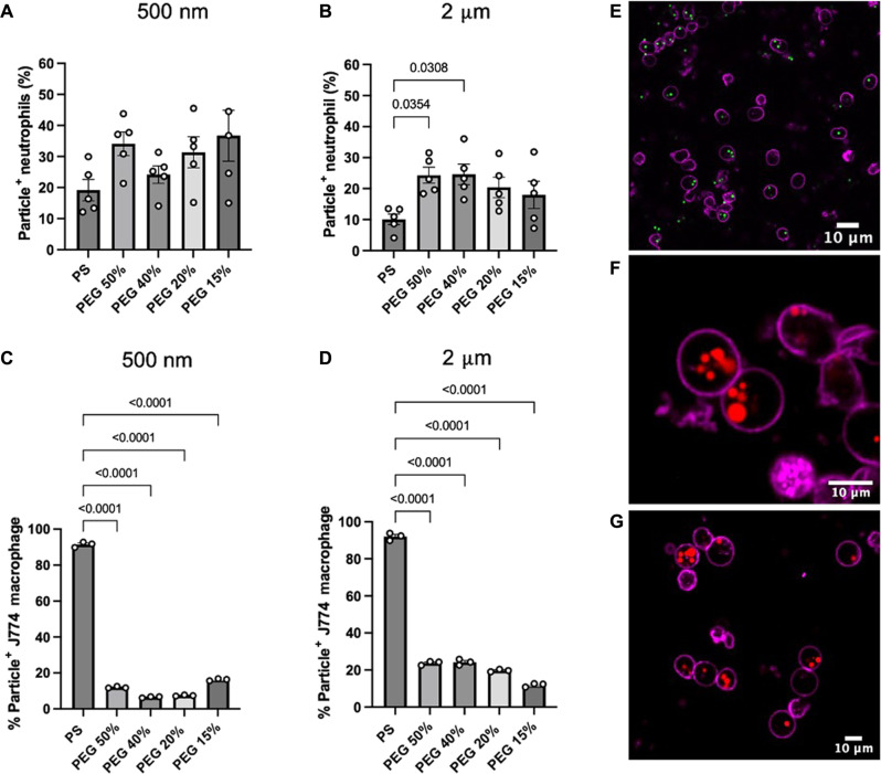
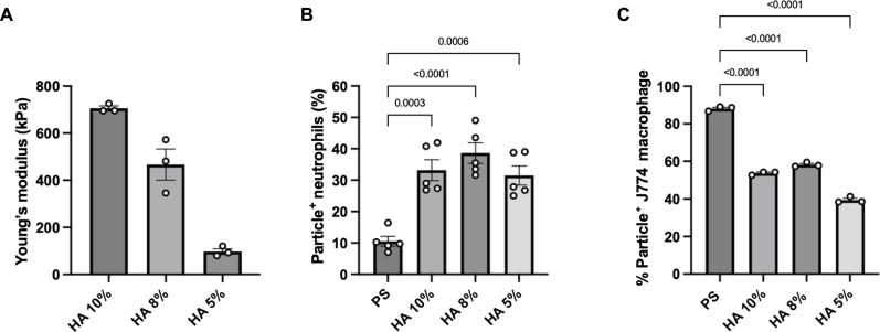
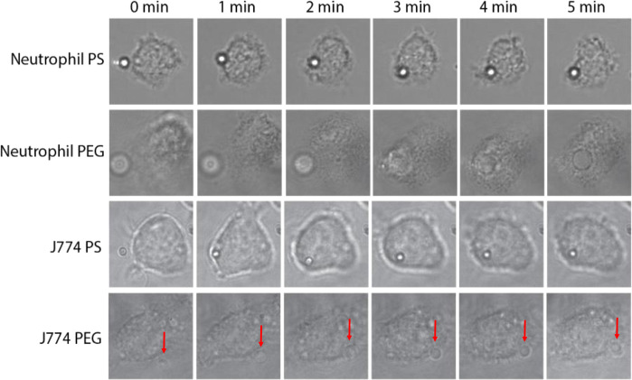
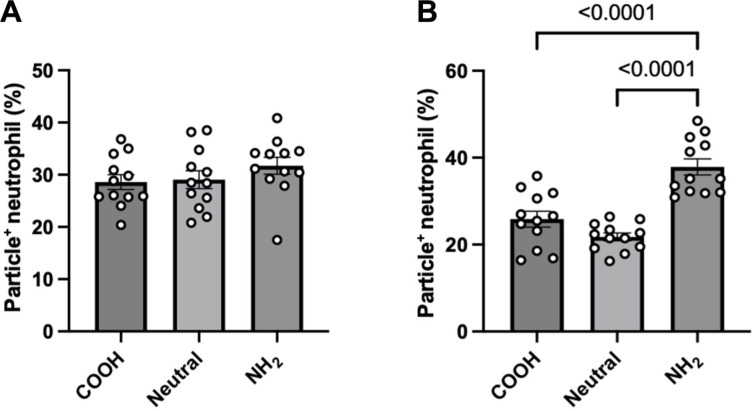
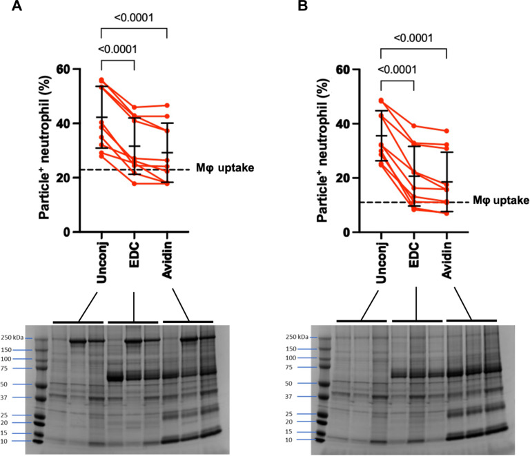
Particle elasticity has widely been established to substantially influence immune cell clearance and circulation time of vascular-targeted carriers (VTCs). However, prior studies have primarily investigated interactions with macrophages, monocytic cell lines, and in vivo murine models. Interactions between particles and human neutrophils remain largely unexplored, although they represent a critical aspect of VTC performance. Here, we explore the impact of particle elasticity on primary human neutrophil phagocytosis using polyethylene glycol-based particles of different elastic moduli. We found that neutrophils effectively phagocytose deformable particles irrespective of their modulus, indicating a departure from established phagocytosis trends seen with other types of immune cells. These findings highlight the observed phenotypic difference between different types of phagocytes and underscore the need to characterize VTC performance using various cell types and animal models that represent human systems closely.


Figures

Fig. 1.
Fig. 1.. Phagocytosis of 2-μm- and 500-nm-sized PS and PEG particles.
Percentage of particle-positive cells by (A and B) primary human neutrophils in whole blood and (C and D) J774 macrophages in Dulbecco’s modified Eagle’s medium (DMEM) with 10% fetal bovine serum (FBS). Samples were incubated with cells for 2 hours before staining, fixing, and analyzing with flow cytometry. At least 5000 events were recorded for each assay. Confocal microscopy images of isolated human neutrophils confirmed that (E) 2-μm PS, (F) 2-μm 50% PEG, (G) and 2-μm 15% PEG particles were internalized by the cells. Statistical analysis was performed using one-way ANOVA with Tukey’s multiple comparisons posttest. For (A) and (B), graph bars show the average particle-positive neutrophils with SEM. Each circle represents an individual blood donor. For (C) and (D), graph bars show the average of three technical replicates of J774 macrophage uptake. For (E) to (G), the scale bar is 10 μm.
Fig. 2.
Fig. 2.. Rheometry and uptake of HA-based hydrogels.
(A) Rheometry of bulk HA hydrogel samples, indicating a wide range of Young’s moduli. Uptake of 2-μm-sized HA-derived spherical particles by (B) primary human neutrophils in whole blood and (C) J774 macrophages in DMEM and 10% FBS medium. Statistics for the phagocytosis assays were performed using one-way analysis of variance (ANOVA) with Tukey’s multiple comparisons test. Bars represent the average percentage of particle-positive cells, and error bars represent SEM with n = 5 blood donors for neutrophils and n = 3 technical replicates for J774 macrophages and rheometry samples.
Fig. 3.
Fig. 3.. Optical tweezer images where a single 2-μm particle was brought into contact with a cell.
Assays were carried out with either primary human neutrophils with PS/PEG or cultured J774 macrophages with PS/PEG. Frames were selected at 1-min intervals to visualize particles as they were engulfed by cells. The PEG particle was unable to be phagocytosed by the J774 macrophage, as indicated by the red arrow near the periphery of the cell.
Fig. 4.
Fig. 4.. Uptake of 2-μm PEG particles with varying zeta potential.
The 2-μm-sized (A) 50% PEG and (B) 15% PEG particles of varying zeta potentials (either positive, negative, or neutral) were incubated with primary human neutrophils in whole blood. Displayed zeta potentials confirmed successful surface modification of surface charge using an amino-group–containing linker. Significance was determined using one-way ANOVA with Tukey’s multiple comparisons test. Error bars represent SEM where n = 12 independent blood donors.
Fig. 5.
Fig. 5.. Uptake of surface-conjugated PEG particles.
The 2-μm-sized (A) 50% and (B) 15% PEG particles either unconjugated (Unconj), activated with EDC, or covalently coated with avidin were incubated with primary human neutrophils in whole blood. The data average is shown as a horizontal check with error bars. The set of three data represent plasma from three distinct donors. For comparison, the dotted line on each graph represents the average uptake of each respective particle type by J774 macrophages at a ratio of 40 particles per cell. The protein coronas of these particles were also visualized using SDS–polyacrylamide gel electrophoresis (PAGE) and stained with Coomassie Blue. There were notable bands for both EDC activation and avidin coating around 60 kDa. Statistics for phagocytosis assays were performed using repeated measures of one-way ANOVA with Dunnett’s multiple comparisons test. Error bars represent SD with n = 10 independent blood donors.
Fig. 6.
Fig. 6.. Biodistribution of PS and 50% PEG particles in BALB/c mice.
(A) Biodistribution of 1.75-μm PS or 2-μm 50% PEG hydrogels after 2 hours intravenous injection of male BALB/c mice. (B) In vivo uptake of PS and PEG particles by mouse neutrophils, quantified as a percentage of particle-positive neutrophils. (C) Particles remaining in circulation 2 hours after injection, quantified as a percentage of particles relative to the number of white blood cells. Statistics were performed using Student’s t test for all plots with n = 5 male mice.

Similar articles

Cited by

References

    1. Fish M. B., Banka A. L., Braunreuther M., Fromen C. A., Kelley W. J., Lee J., Adili R., Holinstat M., Eniola-Adefeso O., Deformable microparticles for shuttling nanoparticles to the vascular wall. Sci. Adv. 7, eabe0143 (2021). - PMC - PubMed
    1. Key J., Palange A. L., Gentile F., Aryal S., Stigliano C., Di Mascolo D., De Rosa E., Cho M., Lee Y., Singh J., Decuzzi P., Soft discoidal polymeric nanoconstructs resist macrophage uptake and enhance vascular targeting in tumors. ACS Nano 9, 11628–11641 (2015). - PubMed
    1. Merkel T. J., Jones S. W., Herlihy K. P., Kersey F. R., Shields A. R., Napier M., Luft J. C., Wu H., Zamboni W. C., Wang A. Z., Bear J. E., DeSimone J. M., Using mechanobiological mimicry of red blood cells to extend circulation times of hydrogel microparticles. PNAS 108, 586–591 (2011). - PMC - PubMed
    1. Merkel T. J., Chen K., Jones S. W., Pandya A. A., Tian S., Napier M. E., Zamboni W. E., DeSimone J. M., The effect of particle size on the biodistribution of low-modulus hydrogel PRINT particles. J. Control. Release 162, 37–44 (2012). - PMC - PubMed
    1. Zhang L., Cao Z., Li Y., Ella-Menye J.-R., Bai T., Jiang S., Softer zwitterionic nanogels for longer circulation and lower splenic accumulation. ACS Nano 6, 6681–6686 (2012). - PubMed

LinkOut - more resources