. 2025 Jan 3;21(1):e1011220.
doi: 10.1371/journal.pgen.1011220. eCollection 2025 Jan.

Multi-modal comparison of molecular programs driving nurse cell death and clearance in Drosophila melanogaster oogenesis

Affiliations

Multi-modal comparison of molecular programs driving nurse cell death and clearance in Drosophila melanogaster oogenesis

Shruthi Bandyadka et al. PLoS Genet. .

Abstract

The death and clearance of nurse cells is a consequential milestone in Drosophila melanogaster oogenesis. In preparation for oviposition, the germline-derived nurse cells bequeath to the developing oocyte all their cytoplasmic contents and undergo programmed cell death. The death of the nurse cells is controlled non-autonomously and is precipitated by epithelial follicle cells of somatic origin acquiring a squamous morphology and acidifying the nurse cells externally. Alternatively, stressors such as starvation can induce the death of nurse cells earlier in mid-oogenesis, manifesting apoptosis signatures, followed by their engulfment by epithelial follicle cells. To identify and contrast the molecular pathways underlying these morphologically and genetically distinct cell death paradigms, both mediated by follicle cells, we compared their genome-wide transcriptional, translational, and secretion profiles before and after differentiating to acquire a phagocytic capability, as well as during well-fed and nutrient-deprived conditions. By coupling the GAL4-UAS system to Translating Ribosome Affinity Purification (TRAP-seq) and proximity labeling (HRP-KDEL) followed by Liquid Chromatography tandem mass-spectrometry, we performed high-throughput screens to identify pathways selectively activated or repressed by follicle cells to employ nurse cell-clearance routines. We also integrated two publicly available single-cell RNAseq atlases of the Drosophila ovary to define the transcriptomic profiles of follicle cells. In this report, we describe the genes and major pathways identified in the screens and the striking consequences to Drosophila melanogaster oogenesis caused by RNAi perturbation of prioritized candidates. To our knowledge, our study is the first of its kind to comprehensively characterize two distinct apoptotic and non-apoptotic cell death paradigms in the same multi-cellular system. Beyond molecular differences in cell death, our investigation may also provide insights into how key systemic trade-offs are made between survival and reproduction when faced with physiological stress.


Conflict of interest statement

The authors have declared that no competing interests exist.

Figures

Fig 1
Fig 1. Nurse cell (NC) clearance is regulated apoptotically or non-apoptotically depending on environmental cues.
(A) Schematic illustrating the different fates of egg chambers. A stage egg 8 chamber is completely surrounded by follicle cells (shown in teal). When the milieu is optimal and sufficient dietary protein is available, oogenesis proceeds (right side). A subset of the follicle cells differentiates into stretch follicle cells (SFCs, blue) and express the phagocytic receptor Draper and the proton pump Vacuolar-ATPase on their plasma membrane, ultimately acidifying the germline sister Nurse Cells (NCs, large black nuclei) externally to clear them. In contrast, when the organism is deprived of nutrition (left side), follicle cells enlarge in place (orange) and engulf the NCs, also by expressing Draper on their surfaces. In both death modalities, FCs clear NCs, albeit using different morphological configurations and biochemical pathways, providing a unique opportunity to study diverse cell death modalities in the same system (B) Venn diagram illustrating that developmental phagoptosis (blue) and starvation-induced death of NCs (orange) share key features such as the expression of Draper on the surface of the follicle cells and the activation of JNK signaling.
Fig 2
Fig 2. Expression of the RpL10Ab-GFP construct for TRAP-seq and the HRP-KDEL construct for LC-MS/MS.
(A-D”) Egg chambers of indicated genotypes stained with DAPI. Merge shows DAPI in cyan and GFP in white. (A-A”) Several ovarioles expressing GR1 > RpL10Ab-GFP in AFCs. Younger egg chambers are located anteriorly (left). Scale bars = 200μ (B-B”) Expression of GR1 > RpL10Ab-GFP in AFCs of mid-stage egg chamber. Scale bars = 50μ (C-C”) Several ovarioles expressing PG150 > RpL10Ab-GFP in SFCs. Scale bars = 200μ (D-D”) Expression of PG150 > RpL10Ab-GFP in SFCs of stage 10 egg chamber. Scale bars = 50μ. (E-F) Egg chambers of indicated genotypes stained for DAPI (cyan) and anti-V5 (magenta) to detect HRP-KDEL. (E) GR1>HRP-KDEL-V5 stage 9 egg chamber shows HRP-KDEL in all follicle cells. (F) PG150>HRP-KDEL-V5 stage 10 egg chamber shows HRP-KDEL in stretch follicle cells only. (G-H) PG150>HRP-KDEL-V5 stage 11 egg chamber with (G) DAPI, (H) biotinylated proteins (Streptavidin-488, green), and (I) HRP-KDEL (anti-V5, magenta). (J) Merge demonstrating that protein biotinylation pattern is restricted to cells expressing HRP-KDEL.
Fig 3
Fig 3. Establishing the translatome, secretome, and transcriptome of AFCs and SFCs.
(A) Schematic illustrating the experimental design for identification of the translatome and secretome from 3 conditions/cell types. GAL4 drivers GR1 and PG150 were used to drive expression of RpL10Ab-GFP in AFCs (fed or protein-starved) and SFCs. The resulting GFP-conjugated ribosomes were immunoprecipitated and the mRNAs bound to the ribosomes were sequenced to produce the translatome. Similarly, to generate the secretome, GR1 and PG150 were used to drive the expression of ss-HRP-KDEL-V5 in AFCs and SFCs. HRP-KDEL expressed in cells localizes to the endoplasmic reticulum and biotinylated when incubated with biotin-phenol and H2O2. The tagged proteins were subsequently isolated using streptavidin beads and analyzed via LC-MS/MS to produce the secretome. (B) PCA Dimension reduction of aggregated gene-level transcript counts in translatome bioreplicates. (C) PCA of peptide intensity values of secretome bioreplicates. (D) UMAP embedding of Seurat RPCA integrated cells from publicly available single-cell RNA-seq atlases of the Drosophila melanogaster ovaries [24,25]. Cells are colored by the dataset of origin. (E) UMAP embedding of the integrated ovary datasets colored by SFC module score. To identify cells most likely to be stretch follicle cells in the integrated dataset, an aggregate score comprising the expression values of canonical SFC markers used in the published individual datasets were computed and projected on to the UMAP. (F) UMAP embedding of the integrated ovary datasets colored by mid/late follicle cell module score. To identify cells most likely to be mid/late follicle cells in the integrated dataset, an aggregate score comprising the expression values of canonical mid/late follicle cell markers used in the published individual datasets were computed and projected on to the UMAP. (G) Cells with a high SFC score (>1) and high mid/late follicle cell score (>2x) were extracted from the integrated dataset and subject to reclustering. Updated PCA and UMAP embeddings were computed for the new mid/late follicle cell and SFC subset and clusters containing mid/late follicle cells and SFCs were identified after normalizing read counts in the new subset.
Fig 4
Fig 4. Functional enrichment of candidates identified in the translatome and secretome reveals pathways common and unique to phagoptosis (Fed SFCs/Fed AFCs) and starvation-death of NCs (Starved AFCs/Fed AFCs).
(A) Scatterplot of differentially translated gene candidates in phagoptosis (Fed SFCs / Fed AFCs–x-axis) and starvation-death (Starved AFCs / Fed AFCs–y-axis) obtained using DESeq2. LFC differentials and FDR adjusted p-values from both analyses were compared to find genes congruently or uniquely regulated in both death modalities. Quadrants 1 and 3 indicate genes congruently up and down-regulated in phagoptosis and starvation-death respectively. Congruency was determined if the direction of LFC was the same and if the adjusted p-value was less than 0.05 in both. (B) Scatterplot of differentially abundant candidates in phagoptosis and starvation-death in the secretome. Proteins common to both death modalities were not found. UniProt identifiers along with gene symbols of detected proteins are specified. (C) Counts of genes regulated congruently and uniquely in phagoptosis and starvation-death of NCs in the translatome. Figure color legend same as in (A). (D) Dot plot of GO-terms enriched in the categories of differentially translated genes identified in (A and C). (E) Dot plot of GO-terms enriched in the secretome of fed SFCs and starved AFCs.
Fig 5
Fig 5. RNAi knockdown of Ca2+ associated cytoskeleton regulators leads to developmental NC clearance defects.
(A-B) Representative images of anterior regions of stage 14 egg chambers from control (GR1 > Luciferase RNAi) and mutant (GR1 > cue RNAi) flies. DNA is labeled with DAPI (cyan). Stage 14 egg chambers are distinguished by two fully developed dorsal appendages (DA), indicated by white arrows. Persisting NC nuclei (PNCN) indicating a disruption to phagoptosis resulting from RNAi are marked by yellow arrows. Scale bars = 50μ. (C) Quantification of PNCN in stage 14 egg chambers. GR1 > cue RNAi demonstrates a strong PNCN phenotype, while the other knockdowns demonstrate a moderate to weak PNCN phenotype. At least 10 flies (20 ovaries) were used for each genotype. (*** p < = 5e-04, **** p < = 5e-05, ns p>0.05, one-sided independent t-test) Raw data available in S5 Table.
Fig 6
Fig 6. The role of innate immune signaling in NC clearance.
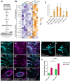
(A) Schematic of Drosophila Toll pathway (B) Heatmap of scaled read counts of NF-κB regulators and antimicrobial peptide genes upregulated by fed SFCs or starved AFCs in the translatome. (C) Quantification of PNCN in stage 14 egg chambers from NF-κB or AMP knockdowns. (*** p< = 5e-04, **** p< = 5e-05, ns p>0.05, one-sided independent t-test). Significance levels are indicated for samples that have at least 3 replicates. (D) Egg chambers from wild-type fly ovaries stained with DAPI (cyan) and anti-Dlg (magenta) to label FC membranes. Scale bars = 200μ. (E) Egg chambers from GR1>SPE RNAi fly ovaries stained with DAPI and anti-Dlg. GR1>SPE RNAi ovarioles contain intact germaria and healthy early egg chambers but show wide-spread degeneration beginning around stage 6. Scale bars = 200μ. (F-H) Images of individual healthy and mid-stage dying egg chambers from wild-type ovaries and a mid-stage dying egg chamber from GR1>SPE RNAi stained with DAPI and anti-Dlg. The GR1>SPE RNAi degenerating egg chamber is lacking most of the follicle cell layer, indicated by white arrows. NC DNA in GR1>SPE RNAi is highly condensed and few are fragmented. Anterior-posterior polarity of the egg chamber is lost, with NCs extending into the posterior end, displacing the oocyte. Scale bars = 50μ. (I) GR1>LexA RNAi starved control shows sporadically degenerating egg chambers, Scale bars = 200μ. (J) GR1>AttD RNAi starved shows increased degeneration of egg chambers Scale bars = 200μ. (K) Quantitative analysis of midstage degenerating egg chambers with one-way ANOVA. (*** p < 0.0008, ns p> 0.1) Graph displays mean + SD with n > 24 females per genotype and condition. Raw data available in S5 Table.
Fig 7
Fig 7. in vivo RNAi screening under fed condition.
(A) Heatmap of scaled read counts of major signaling pathway genes that are significantly differentially regulated in SFCs. (B,C) GR1>Dad RNAi and GR1>LBR RNAi result in a “dumpless” phenotype (outlined in magenta), in addition to increased mid-stage death (orange arrows). Scale bar = 100μ. (D) GR1>numb RNAi results in mid-stage death accompanied by abnormal egg chamber morphology (yellow arrows). Scale bar = 100μ. (E) GR1 > prosα3 RNAi ovaries have a strong PNCN phenotype. Scale bar = 50μ. (F) NC nuclei in a stage 9 egg chamber from CantonS ovary. Scale bar = 50μ. (F’, F”) 3D surface plot of individual nuclei in stage 9 egg chamber from CantonS ovary after z-projection (max-intensity across stacks). Colors of peaks represent pixel intensity across z-stacks, with darker colors representing lower pixel intensity in the area (low DAPI staining) and lighter colors representing higher pixel intensity (robust DAPI staining). (G) NC nuclei in a stage 9 egg chamber from GR1 > wat RNAi ovary. Scale bar = 50μ. (G’, G”) 3D surface plot of individual nuclei in stage 9 egg chamber from GR1 > wat RNAi ovary after z-projection (max-intensity across stacks) reveals increased vacuolization. (H) Quantification of PNCN in stage 14 egg chambers of select candidates from Table 1. (* p < 5e-02, ** p < = 5e-03, *** p < = 5e-04, **** p < = 5e-05, ns p>0.05, one-sided independent t-test). Error bars indicate mean + S.D. across at least 3 replicates, with each replicate having at least 5 flies. Raw data available in S5 Table.

References

    1. Lebo DPV, McCall K. Murder on the Ovarian Express: A Tale of Non-Autonomous Cell Death in the Drosophila Ovary. Cells. 2021;10. doi: 10.3390/cells10061454 - DOI - PMC - PubMed
    1. King RC. Ovarian Development in Drosophila Melanogaster. Academic Press: New York, NY, USA, ISBN 9780124081505. 1970. [cited 20 Dec 2023]. Available: https://cir.nii.ac.jp/crid/1370004237568899203.
    1. Spradling A. Developmental genetics of oogenesis. The development of Drosophila melanogaster. 1993. Available: https://ci.nii.ac.jp/naid/10016991326/
    1. Giorgi F, Deri P. Cell death in ovarian chambers of Drosophila melanogaster. J Embryol Exp Morphol. 1976;35: 521–533. - PubMed
    1. Drummond-Barbosa D, Spradling AC. Stem cells and their progeny respond to nutritional changes during Drosophila oogenesis. Dev Biol. 2001;231: 265–278. - PubMed

Substances

LinkOut - more resources