. 2025 Feb 6;112(2):353-373.
doi: 10.1016/j.ajhg.2024.12.006. Epub 2025 Jan 2.

CIROZ is dispensable in ancestral vertebrates but essential for left-right patterning in humans

Affiliations

CIROZ is dispensable in ancestral vertebrates but essential for left-right patterning in humans

Emmanuelle Szenker-Ravi et al. Am J Hum Genet. .

Abstract

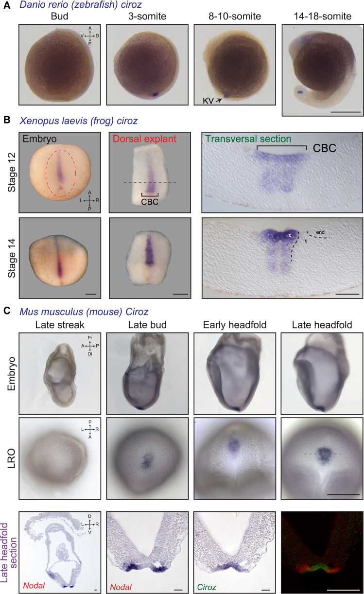
Four genes-DAND5, PKD1L1, MMP21, and CIROP-form a genetic module that has specifically evolved in vertebrate species that harbor motile cilia in their left-right organizer (LRO). We find here that CIROZ (previously known as C1orf127) is also specifically expressed in the LRO of mice, frogs, and fish, where it encodes a protein with a signal peptide followed by 3 zona pellucida N domains, consistent with extracellular localization. We report 16 individuals from 10 families with bi-allelic CIROZ inactivation variants, which cause heterotaxy with congenital heart defects. While the knockout of Ciroz in mice also leads to situs anomalies, we unexpectedly find that its targeted inactivation in zebrafish and Xenopus does not lead to observable LR anomalies. Moreover, CIROZ is absent or obsolete in select animals with motile cilia at their LRO, including Carnivora, Atherinomorpha fish, or jawless vertebrates. In summary, this evo-devo study identifies CIROZ as an essential gene for breaking bilateral embryonic symmetry in humans and mice, whereas we witness its contemporary pseudogenization in discrete vertebrate species.

Keywords: C1orf127; CIROP; CIROZ; DAND5; MMP21; PKD1L1; ZP-N; ZP2; evo-devo; gene loss; heterotaxy; laterality; left-right organizer; motile cilia; pseudogenization.


Conflict of interest statement

Declaration of interests The authors declare that they have no competing interests.

Figures

None
Graphical abstract
Figure 1
Figure 1
Pseudogenization of CIROZ in laurasiatherian mammals, reptiles, and birds (A) Simplified evolutionary tree of vertebrate species. The dashed red line indicates that the absence of motile cilia at the LRO has not yet been shown experimentally in cetaceans, as opposed to birds (chicken), reptiles (gecko), and Artiodactyla (pig). The red lines indicate where the CIROZ gene has been lost, the purple lines where it has accumulated deleterious mutations during evolution, and the blue lines where it encodes a shorter protein. Informed consent from the parent was obtained to publish the photograph of the toddler. (B) CIROZ schematic structures in human (Homo sapiens [h]), mouse (Mus musculus [m]), cat (Felis catus [c]), whale (Balaenoptera acutorostrata scammoni [w]), camel (Camelus bactrianus [cam]), Xenopus laevis (xl), Xenopus tropicalis (xt), and zebrafish (Danio rerio [zf]). Red lines indicate premature stop or frameshift variants. (C) Representation of the CIROZ locus in representative jawed vertebrates whose genomes have been sequenced, showing the loss of the gene in turtles (Reptilia) as well as in medaka (fish). Block arrows represent genes, with the direction of arrows denoting transcriptional orientation. Orthologous genes are shown with the same color.
Figure 2
Figure 2
Identification of bi-allelic variants in CIROZ in individuals with HTX (A) Pedigrees of 10 families with individuals presenting with heterotaxy (HTX). The CIROZ genotypes for available individuals are indicated. Double lines between symbols indicate consanguineous marriages. Squares, circles, diamonds, and triangles denote males, females, unknown gender individuals, and fetuses, respectively. Open and filled symbols are used for unaffected and affected family members, respectively, and deceased individuals are indicated by a diagonal slash through the symbol. Small black circles indicated spontaneous abortion. The gray question mark indicates a denied consanguinity in family 8. TOP, termination of pregnancy; wt, wild type; mut, mutation. (B) Genomic and protein schematic structures of human CIROZ. The position and nature of the identified variants are indicated. CIROZ known protein domains are highlighted: signal peptide (SP; yellow) and DUF44556 (domain of unknown function, orange). (C) Comparison of the heart phenotype in CIROZ-deficient individuals compared to affected individuals with causative variants in one of the 4 other HTX genes of the module (MMP21, CIROP, PKD1L1, and DAND5). CHDs, congenital heart defects. Only individuals with a confirmed CIROZmut/mut genotype were included in this analysis (n = 16). (D) Comparison of the situs clinical outcome (situs solitus, situs ambiguus, or situs inversus) in individuals with bi-allelic loss-of-function variants in any of the 5 genes of the module (MMP21, CIROP, PKD1L1, DAND5, and CIROZ) or that of 6 genes (CCDC39, CCDC40, DNAH5, DNAH11, DNAI1, and DNAI2) whose bi-allelic mutation leads to primary ciliary dyskinesia (CILD).
Figure 3
Figure 3
CIROZ is expressed in the LRO of zebrafish, Xenopus, and mouse embryos (A) Whole-mount in situ hybridization (WISH) of ciroz in Danio rerio (zebrafish) embryos at indicated stages. The black arrow points to the expression in the Kupffer’s vesicle (KV). Scale bar, 0.25 mm. Similar results were obtained in at least three independent experiments with 50 embryos each. (B) WISH of ciroz in Xenopus laevis (frog) embryos at indicated stages. Colored dotted lines indicate the respective sections. Scale bar, 0.2 mm. c, central-flow-generating LRO cells; s, sensory LRO cells; end, lateral endodermal cells. Similar results were obtained in three independent experiments with at least 15 embryos each. (C) WISH of Ciroz in Mus musculus (mouse) embryos at indicated stages. Representative images of Ciroz expression are shown (n = 3 embryos per stage were stained). Whole embryos and zoomed-in pictures of the LRO are shown. Section in situ hybridization (SISH) of Nodal and Ciroz were performed on serial sections (purple dashed line) of late headfold embryos. Representative images are shown (n = 3 embryos). Section of a whole embryo with Nodal staining and zoomed-in pictures of the LRO for Nodal and a neighboring section for Ciroz are shown. The overlap image shows a false color merge of the picture of Nodal switched to red and Ciroz switched to green. Scale bars, 0.2 mm. V, ventral; D, dorsal; A, anterior; P, posterior; L, left; R, right; CBC, circumblastoporal collar; Pr, proximal; Di, distal; LRO, left-right organizer.
Figure 4
Figure 4
Xenopus crispants or morphants for ciroz do not exhibit LR defects (A) Depiction of genomic and protein structures of human and Xenopus laevis Ciroz. The Xenopus laevis sequence used is Genbank: XM_041569138.1/XP_041425072. Ciroz protein domains are highlighted: signal peptide (SP; yellow) and DUF44556 (domain of unknown function, orange). The sites targeted by CRISPR gRNA1 and gRNA2 are indicated by two red pairs of scissors in introns 2 and 11, respectively. The sites targeted by the translation-blocking (TBMOs; purple) and splice-blocking (SBMOs; green) morpholinos are also indicated. h, human; xl, Xenopus laevis. (B) Schematic representation of secretion assay using dissociated and cultured animal caps previously microinjected at the 8-cell stage with mRNAs encoding CIROZ-HA, CIROP, or MMP21-FLAG. Western blot performed on the spent supernatant reveals that each of these 3 proteins are readily secreted, consistent with their conserved N-terminal SP. (C) Scoring of internal organs of stage 45 Xenopus larvae. Situs solitus represents the normal situation where the heart is on the left, the gallbladder is on the right, and the intestine rotation is anticlockwise. Situs inversus is the complete mirror image of situs solitus, and situs ambiguus represents any situation in between. n = total number of embryos analyzed from at least 3 independent experiments. Data are mean ± SEM. NS, not significant. Two-way ANOVA with Tukey's test for multiple comparisons (normal condition, situs solitus). wt, wild type. (D) Absence of external phenotype at stage 45. Scale bar, 1 mm.
Figure 5
Figure 5
Zebrafish without Ciroz do not exhibit LR defects (A) Depiction of genomic and protein structures of human and zebrafish Ciroz. The zebrafish sequence used is Genbank: XM_009296969/XP_009295244 using the alternate exon 1 (ex1) described in Figure S5. Ciroz protein domains are highlighted: signal peptide (SP; yellow) and DUF44556 (domain of unknown function, orange). The site targeted by the CRISPR gRNA is indicated by the red pair of scissors in exon 6. Three different alleles, lof1 (red), lof2 (purple), and lof3 (green), were obtained, all leading to a frameshift (black) with an early stop codon. aa, amino acid; h, human; lof, loss-of-function; zf, zebrafish; wt, wild type. (B) Left: qPCR for ciroz relative to actin at indicated stages. Data are mean ± SEM. ∗∗p = 0.0016 and ∗∗∗∗p < 0.0001. One-way ANOVA with Tukey’s test for multiple comparisons. n = at least 3 biological replicates of 30–50 embryos. dpf, days postfertilization. Right: whole-mount ciroz in situ hybridization in zebrafish embryos from indicated crosses at the 4-somite stage. Zooms on the Kupffer’s vesicles (KV) are shown. Black arrow points to expression in the KV of control embryos. The number of analyzed embryos is indicated. Scale bar, 0.25 mm. (C) Scoring of cardiac looping at 48 hpf of embryos from indicated crosses. Genotypes of the parents and percentage of ciroz−/− embryos are indicated. Normal, no, or inverted looping is classified in white, light orange, or dark orange, respectively. n = total number of embryos analyzed from at least 3 independent experiments. Data are mean ± SEM. NS, not significant. Two-way ANOVA with Tukey’s test for multiple comparisons (normal looping condition). Z, zygotic; MZ, maternal zygotic. (D) Scoring of liver (red) and pancreas (green) positions using the LiPan transgenic line in embryos from indicated crosses. Genotypes of the parents and percentage of ciroz−/− embryos are indicated. Normal position (liver on the left and pancreas on the right) and inverted position are classified in white and pink, respectively. n = total number of embryos analyzed from at least 3 independent experiments. Data are mean ± SEM. NS, not significant. Two-way ANOVA with Tukey’s test for multiple comparisons (normal condition). Images are side views of embryos (right or left as indicated). P, posterior; A, anterior. (E) Scoring of stronger lov expression side at 72 hpf of embryos from indicated crosses. Genotypes of the parents and percentage of ciroz−/− embryos are indicated. Left (normal), right, or equal are classified in white, dark green, and light green, respectively. n = total number of embryos analyzed from at least 3 independent experiments. Data are mean ± SEM. NS, not significant. Two-way ANOVA with Tukey’s test for multiple comparisons (normal condition). (F) Scoring of lefty2 expression side at 24 hpf of embryos from indicated crosses. Genotypes of the parents and percentage of ciroz−/− embryos are indicated. Left (normal), right, bilateral, or absent are classified in white, dark blue, light blue, or black, respectively. n = total number of embryos analyzed from at least 3 independent experiments. Data are mean ± SEM. NS, not significant. Two-way ANOVA with Tukey’s test for multiple comparisons (normal condition). (G) Absence of external phenotype at 72 hpf. Z, zygotic; MZ, maternal zygotic. Scale bar, 1 mm.
Figure 6
Figure 6
The N-terminal half of CIROZ bears homology to the 3 ZP-N domains found in ZP2 (A) Protein schematic structures of human CIROZ and human ZP2. The two proteins share the following domains: a signal peptide (SP; yellow), a zona pellucida N-terminal domain 1 (ZP-N1; green), ZP-N2 (cyan), and ZP-N3 (purple). ZP2 also contains a ZP module (ZP-N + ZP-C, red), internal and external hydrophobic patches (IHPs and EHPs, respectively; brown), a conserved furin cleavage site (CFCS; blue), and a transmembrane domain (TM; black). Purple vertical lines: conserved cysteines; known (full purple lines) and predicted (dashed purple lines) cysteine bonds are shown on top. (B) AlphaFold protein structure prediction for mouse CIROZ and mouse ZP2. The domains are indicated with the same color code as in (A). Insets: comparison of the ZP-N1 domain of CIROZ (AlphaFold2) to that of ZP2 (PDB: 5II6), highlighting the 4 conserved cysteines (purple). (C) Anti-HA Western blot analysis on conditioned media from HEK293T cells transfected with indicated human CIROZ-HA constructs. m, monomer; d, dimer; t, trimer; SDS, sodium dodecyl sulfate; DTT, dithiothreitol.

References

    1. Blum M., Andre P., Muders K., Schweickert A., Fischer A., Bitzer E., Bogusch S., Beyer T., van Straaten H.W.M., Viebahn C. Ciliation and gene expression distinguish between node and posterior notochord in the mammalian embryo. Differentiation. 2007;75:133–146. - PubMed
    1. Essner J.J., Amack J.D., Nyholm M.K., Harris E.B., Yost H.J. Kupffer’s vesicle is a ciliated organ of asymmetry in the zebrafish embryo that initiates left-right development of the brain, heart and gut. Development. 2005;132:1247–1260. - PubMed
    1. Nonaka S., Tanaka Y., Okada Y., Takeda S., Harada A., Kanai Y., Kido M., Hirokawa N. Randomization of left-right asymmetry due to loss of nodal cilia generating leftward flow of extraembryonic fluid in mice lacking KIF3B motor protein. Cell. 1998;95:829–837. - PubMed
    1. Nonaka S., Shiratori H., Saijoh Y., Hamada H. Determination of left-right patterning of the mouse embryo by artificial nodal flow. Nature. 2002;418:96–99. - PubMed
    1. Hamada H. Molecular and cellular basis of left-right asymmetry in vertebrates. Proc. Jpn. Acad. Ser. B Phys. Biol. Sci. 2020;96:273–296. - PMC - PubMed