

Radiological markers of CSF α-synuclein aggregation in Parkinson's disease patients
- PMID: 39753572
- PMCID: PMC11698941
- DOI: 10.1038/s41531-024-00854-4
Radiological markers of CSF α-synuclein aggregation in Parkinson's disease patients
Abstract
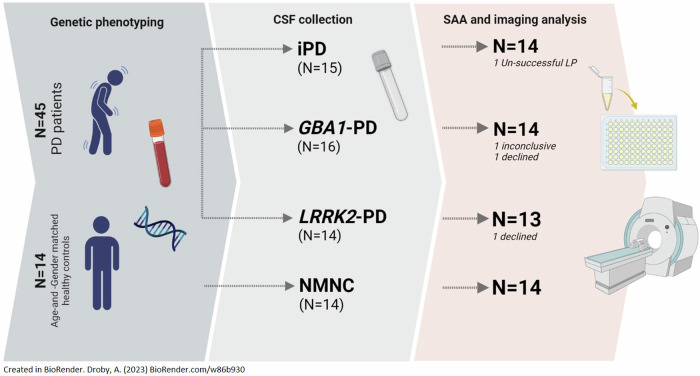
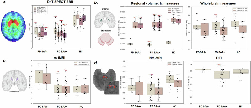
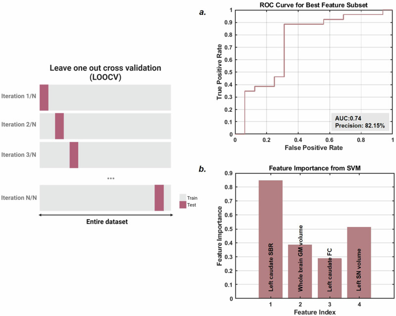
Alpha-synuclein (αS) aggregation is a widely regarded hallmark of Parkinson's disease (PD) and can be detected through synuclein amplification assays (SAA). This study investigated the association between cerebrospinal fluid (CSF) radiological measures in 41 PD patients (14 iPD, 14 GBA1-PD, 13 LRRK2-PD) and 14 age-and-sex-matched healthy controls. Quantitative measures including striatal binding ratios (SBR), whole-brain and deep gray matter volumes, neuromelanin-MRI (NM-MRI), functional connectivity (FC), and white matter (WM) diffusion-tensor imaging (DTI) were calculated. Nine LRRK2-PD patients were SAA-negative (PD-SAA-). PD-SAA+ patients showed lower whole-brain gray matter, putamenal, brainstem, and substantia nigra volumes, reduced FC in the left caudate, and lower fractional anisotropy in the left fronto-occipital fasciculus compared to PD-SAA-. Taken together, αS aggregation was observed in iPD, GBA1-PD, and 38% of LRRK2-PD patients, and this was associated with reduced regional brain volumes, altered caudal FC, and SBRs. These changes were less pronounced in PD-SAA-, possibly suggesting a milder neurodegenerative process.
© 2025. The Author(s).
Conflict of interest statement
Competing interests: AD is an Associate Editor for npj Parkinson’s Disease and receives honoria from Springer Nature. He was not involved in the journal’s review of, or decisions related to, this manuscript. AY-V, BC, NO, AB-S, MG-W, OG, and AO-U report no competing interests. KBF, OSM, and JCS are employees of and own stock in Biogen. RNA research is funded by the Michael J. Fox Foundation (MJFF) and the Parkinson’s Foundation. He received consultation fees from Genzyme/Sanofi, Takeda, and Gain Therapeutics. JMC is a former employee of Biogen, and reports no competing interests relevant to this work. NG has no competing interests pertaining to this work. He serves as a member of the editorial board for the Journal of Parkinson’s Disease. He serves as a consultant to Sionara, Accelmed, Teva, NeuroDerm, Intec Pharma, Pharma2B, Denali, and Abbvie. He received royalties from Lysosomal Therapeutics (LTI) and payment for lectures at Teva, UCB, Abbvie, Sanofi-Genzyme, Bial and Movement Disorder Society. He received research support from the Michael J Fox Foundation, the National Parkinson Foundation, the European Union 7th Framework Program, and the Israel Science Foundation, as well as from the Teva NNE program, Biogen, LTI, and Pfizer. AM reports no competing interests relevant to this work. AM is an Associate Editor for npj Parkinson’s Disease and receives Honoria from Springer Nature. She was not involved in the journal’s review of, or decisions related to, this manuscript. She reports serving as an advisor to Neuroderm. AT reports receiving honoraria from Abbvie, research funding from MJFF, and consultation fees from Capsida Inc. He reports no competing interests relevant to this work.
Figures
References
-
- Dickson, D. W. et al. Neuropathological assessment of Parkinson’s disease: refining the diagnostic criteria. Lancet Neurol.8, 1150–1157 (2009). - PubMed
-
- Atarashi, R. et al. Ultrasensitive human prion detection in cerebrospinal fluid by real-time quaking-induced conversion. Nat. Med.17, 175–178 (2011). - PubMed
-
- Gan-Or, Z. et al. LRRK2 and GBA mutations differentially affect the initial presentation of Parkinson disease. Neurogenetics11, 121–125 (2010). - PubMed
-
- Goldstein, O. et al. Revisiting the non-Gaucher-GBA-E326K carrier state: Is it sufficient to increase Parkinson’s disease risk? Mol. Genet. Metab.128, 470–475 (2019). - PubMed
LinkOut - more resources
Full Text Sources
