. 2025 Jan 3;15(1):693.
doi: 10.1038/s41598-024-84437-6.

hsa-mir-483-3p modulates delayed breast cancer recurrence

Affiliations

hsa-mir-483-3p modulates delayed breast cancer recurrence

Ok-Hyeon Kim et al. Sci Rep. .

Erratum in

Abstract

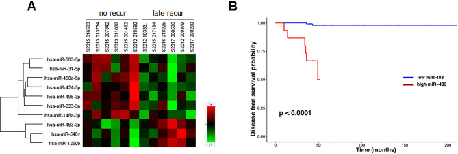
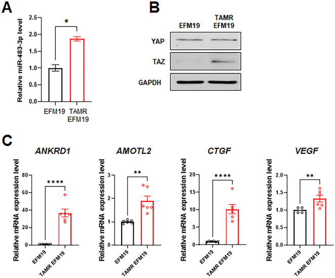
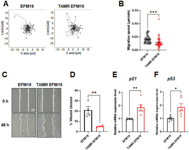
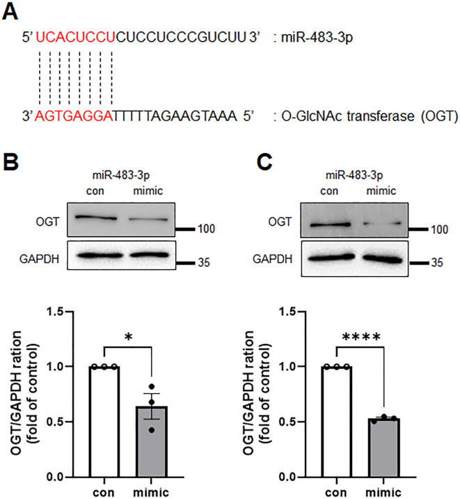
Patients with estrogen receptor-positive breast cancer undergoing continuous adjuvant hormone therapy often experience delayed recurrence with tamoxifen use, potentially causing adverse effects. However, the lack of biomarkers hampers patient selection for extended endocrine therapy. This study aimed to elucidate the molecular mechanisms underlying delayed recurrence and identify biomarkers. When miRNA expression was assessed in luminal breast cancer tissues with and without delayed recurrence using NanoString, a significant increase in the expression of miR483-3p was observed in samples from patients with delayed recurrence compared with those without. miR483-3p expression was elevated in tamoxifen resistant (TAMR) EFM19 cells than in non-resistant EFM19 cells. Notably, genes associated with cancer metastasis (AMOTL2, ANKRD1, CTGF, and VEGF) were upregulated in TAMR EFM19 cells, although cell motility and proliferation were reduced. Transfection of miR483-3p mimics into both non-resistant EFM19 and MCF7 cells resulted in increased expression of cancer metastasis-related genes, but decreased proliferation and migration. Given that miR483-3p can bind to the 3'UTR region of O-GlcNAc transferase (OGT) and potentially affect its protein expression, we examined OGT protein levels and found that transfection with miR483-3p mimics selectively reduced OGT expression. Overall, breast cancer cells subjected to long-term hormone therapy displayed elevated miR483-3p expression, reducing motility and dormancy induction via decreased OGT expression. These findings suggest that miR483-3p is a potential biomarker for long-term endocrine therapy.

Keywords: Breast cancer; Delayed recurrence; Dormancy; miR483-3p.


Conflict of interest statement

Declarations. Competing interests: The authors declare no competing interests. Ethics approval and consent to participate: For human samples, approval was obtained from the Institutional Review Board of Chung-Ang University Hospital (IRB number 2301-019-539). A waiver of informed consent was granted by the Institutional Review Board of Chung-Ang University Hospital.

Figures

Fig. 1
Fig. 1
miRNA screening from patient samples with delayed recurrence or those without. (A) Heat map showing the NanoString gene expression profiles of patient samples with delayed recurrence or those without. (B) Kaplan–Meier survival curve for miR483-3p associated with overall survival in breast cancer. The x-axis represents overall survival time (months) and the y-axis represents survival function.
Fig. 2
Fig. 2
miR-483-3p and YAP/TAZ signaling appeared enhanced in TAMR breast cancer cell. (A) Quantitative RT-PCR results showing the relative indicated miRNA levels in parental EFM19 and TAMR EFM19 cells (unpaired t-test, **p < 0.01; n = 3). (B) YAP and TAZ protein levels in parental EFM19 and TAMR EFM19 cells were evaluated using immunoblotting. (C) Quantitative RT-PCR results indicating relative mRNA levels in parental EFM19 and TAMR EFM19 cells (unpaired t-test, **p < 0.01, ****p < 0.0001; n = 3).
Fig. 3
Fig. 3
TAMR EFM19 cells show reduced motility and dormancy in related gene expression. (A) Spatial tracking of parental or TAMR EFM19 cell movement during the 6 h time-lapse imaging where each cell lies at the origin (0,0) at t = 0 h. The plots show the motility of individual cells in one representative experiment. Cells are indicated as black dots. The movement of each cell was traced using ImageJ software with a manual tracking plugin. The migration speed graphs were summarized using n = 50 individual cell tracking results. (B) Quantification of migration speed revealed the cellular velocities of individual cells (n = 3 independent experiments; unpaired t-test, ***p < 0.001). (C) Wound healing ability in parental EFM19 and TAMR EFM19 cells. Photos were captured at 0 h and 48 h. The closed area indicated by the white line was quantified using ImageJ software. Scale bar = 200 μm. (D) Graph shows the percentage wound closure as quantified using ImageJ software (n = 4; unpaired t-test, **p < 0.01). (E,F) Gene expression levels of p53 and p21 in parental and TAMR EFM19 cells were evaluated by quantitative real-time reverse transcription polymerase chain reaction experiments (qRT-PCR; n = 3 independent experiment, unpaired t-test, *p < 0.05, **p < 0.01).
Fig. 4
Fig. 4
miR483-3p mimics dormancy-like features of breast cancer cells. (A) The expression level of miR483-3p was evaluated using qRT-PCR in parental EFM19 cells after transfection with miRNA control or miR483-3p mimic (unpaired t-test, ****p < 0.0001, n = 3). (B) and (C) Parental EFM19 cells transfected with miRNA control or miR483-3p mimic were evaluated for their proliferation rate at 12 h up to 48 h. Cell viability was assessed using the alamarBlue assay (Two-way ANOVA, *p < 0.05, **p < 0.01, ***p < 0.001, and ****p < 0.0001). (D) Gene expression levels of AMOTL2, ANRKD1, CTGF, PLOD2, and KRT80 in parental EFM19 cells transfected with miRNA control or miR483-3p mimic were evaluated using qRT-PCR (n = 3 independent experiments, unpaired t-test, *p < 0.05, **p < 0.01, ****p < 0.0001). Wound healing ability of parental MCF7 (E) and EFM19 (F) cells transfected with miRNA control or miR483-3p mimic. Photos were taken at 0 h and 48 h. The closed area indicated with a white line was quantified using ImageJ software. Scale bar = 100 μm. Graphs show the calculated wound closure percentage as mentioned in the Methods section (unpaired t-test, *p < 0.05, **p < 0.01).
Fig. 5
Fig. 5
miR483-3p interrupts the expression of OGT. (A) miR483-3p binding sites in the 3’UTR of OGT based on TargetScan prediction. Immunoblot analysis was conducted on parental MCF7 (B) and EFM19 (C) cells transfected with miRNA control or miR483-3p. Graph shows the quantification of the protein expression levels of OGT compared with GAPDH (n = 3, unpaired t-test, *p < 0.05, ****p < 0.0001).

References

    1. Demicheli, R. et al. Recurrence and mortality according to estrogen receptor status for breast cancer patients undergoing conservative surgery. Ipsilateral breast tumour recurrence dynamics provides clues for tumour biology within the residual breast. BMC Cancer. 10, 656 (2010). - PMC - PubMed
    1. Davies, C. et al. Long-term effects of continuing adjuvant tamoxifen to 10 years versus stopping at 5 years after diagnosis of oestrogen receptor-positive breast cancer: ATLAS, a randomised trial. Lancet381, 805–816 (2013). - PMC - PubMed
    1. Ribnikar, D., Sousa, B., Cufer, T. & Cardoso, F. Extended adjuvant endocrine therapy - A standard to all or some? Breast32, 112–118 (2017). - PubMed
    1. Park, Y. H. et al. Pan-asian adapted ESMO Clinical Practice guidelines for the management of patients with early breast cancer: A KSMO-ESMO initiative endorsed by CSCO, ISMPO, JSMO, MOS, SSO and TOS. Ann. Oncol.31, 451–469 (2020). - PubMed
    1. Burstein, H. J. et al. Adjuvant endocrine therapy for women with hormone receptor-positive breast cancer: American Society of Clinical Oncology Clinical Practice Guideline focused update. J. Clin. Oncol.32, 2255–2269 (2014). - PMC - PubMed

Publication types

MeSH terms