. 2025 Jan 3;15(1):625.
doi: 10.1038/s41598-024-84965-1.

Development of the membrane ceiling method for in vitro spermatogenesis

Affiliations

Development of the membrane ceiling method for in vitro spermatogenesis

Maki Kamoshita et al. Sci Rep. .

Abstract

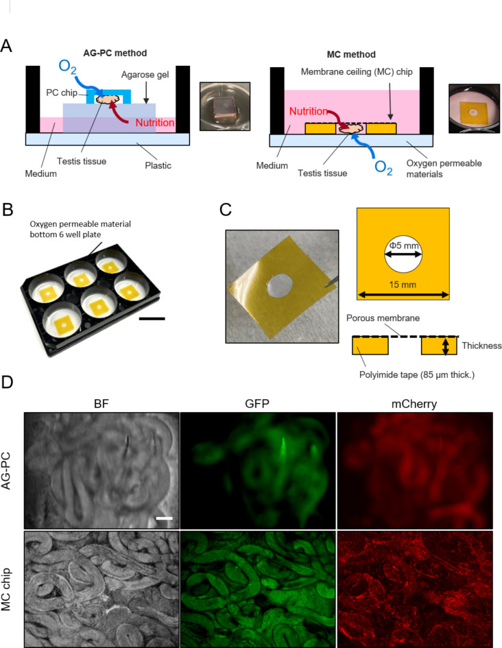
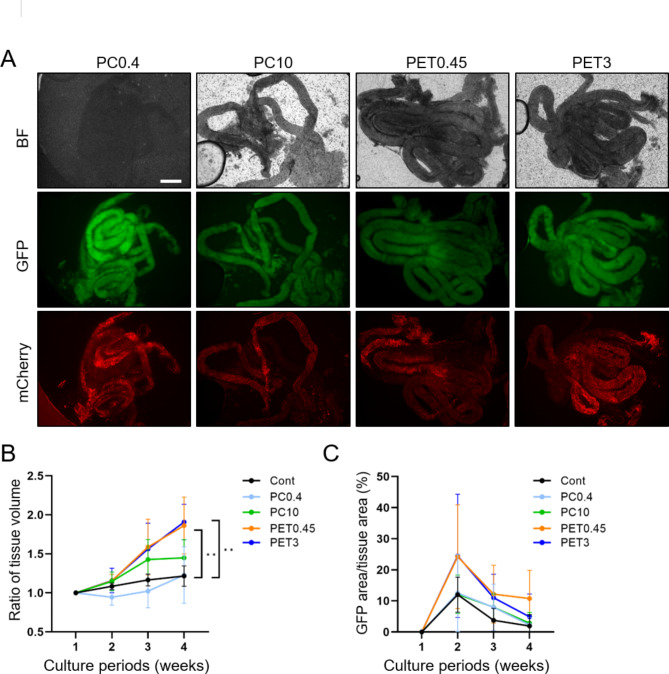
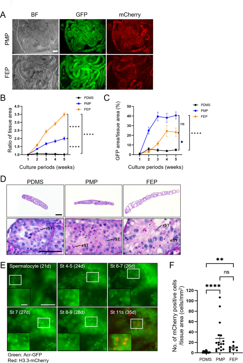
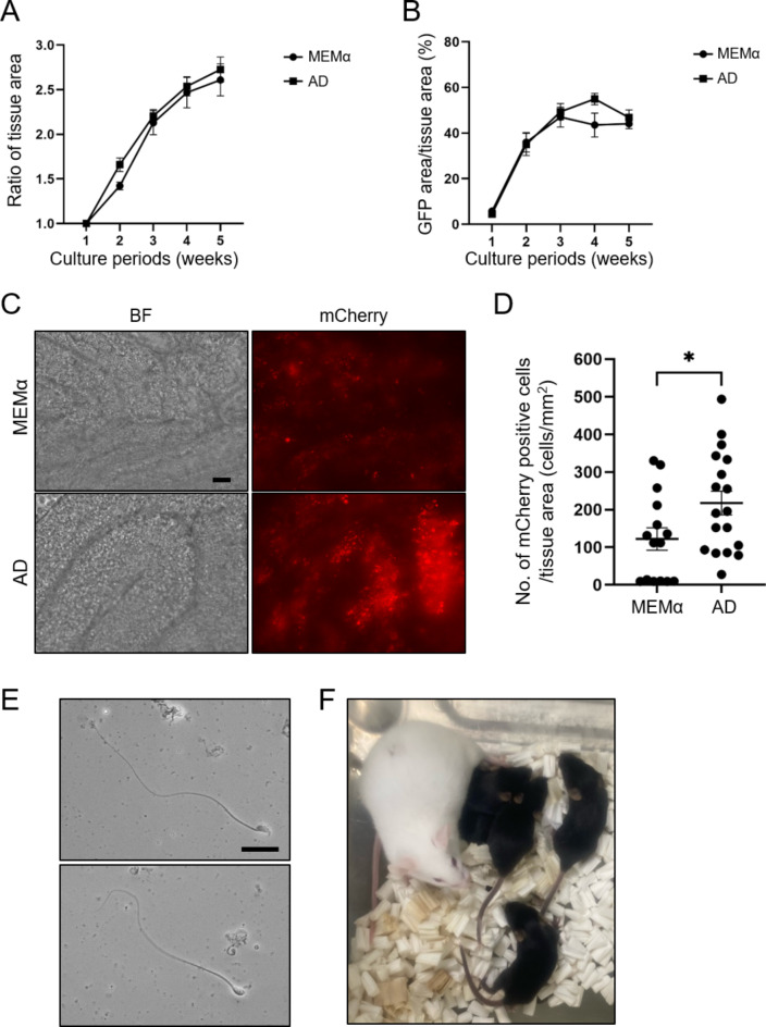
Spermatogenesis is one of the most complex processes of cell differentiation and its failure is a major cause of male infertility. Therefore, a proper model that recapitulates spermatogenesis in vitro has been long sought out for basic and clinical research. Testis organ culture using the gas-liquid interphase method has been shown to support spermatogenesis in mice and rats. However, the conventional method using agarose gel has limitations including medium replacement efficiency and live imaging because agarose absorbs medium and is not transparent. To overcome this issue, we developed a new device using microporous membranes and oxygen-permeable materials. Mouse testes sandwiched between a microporous polyethylene terephthalate (PET) membrane on top and an oxygen-permeable 4-polymethyl-1-pentene polymer (PMP) membrane base maintained spermatogenesis over months. The chamber volume was minimized to 0.1% of the culture medium. Weekly time-lapse live imaging enabled us to observe transgenically fluorescent acrosome and nuclear shape formation throughout spermatogenesis. Finally, we obtained healthy fertile offspring from spermatozoa generated in our system. The device could be used not only for basic research to understand spermatogenesis but also for applied research, such as diagnosing and treating male infertility.

Keywords: 4-Polymethyl-1-pentene polymer (PMP); Fluorinated ethylene-propylene copolymer (FEP); In vitro spermatogenesis; Membrane ceiling chip; Polydimethylsiloxane (PDMS); Testis culture.


Conflict of interest statement

Declarations. Competing interests: Authors K.E., J.Y., and S.Y. were employed by Mitsui Chemicals, Inc. The remaining authors declare that the research was conducted in the absence of any commercial or financial relationships that could be construed as a potential conflict of interest.

Figures

Fig. 1
Fig. 1
Device design. (A) Schematic illustrations and pictures of the conventional method using agarose gel and PDMS ceiling chip (AG-PC) (left) and our novel method consisting of the membrane ceiling (MC) chip and an oxygen-permeable material (MC method) (right). (B) Picture of the MC method. Scale bar = 25 mm. (C) Picture and schematic illustrations of the MC chip. (D) Representative images of testis tissue in the AG-PC and MC method observed by an inverted microscope (BZ-X700). PDMS was used to line the base of the well plate. Scale bar = 100 μm.
Fig. 2
Fig. 2
Optimization of porous membranes used for the MC chip. (A) Representative images using an inverted microscope (BZ-X700) of the MC chip made of various porous membranes as indicated. Scale bar = 0.5 mm. (B) The ratio of tissue volume expansion and (C) the GFP-positive rate over 4 weeks with MC chips consisting of indicated membranes. **P < 0.01 and P > 0.05 if no indication. Twelve (Cont), 8 (PC0.4), 8 (PC10), 8 (PET0.45), and 8 (PET3) tissues were used for the experiments. Images were taken by IX73 for quantifications.
Fig. 3
Fig. 3
Optimization of oxygen-permeable materials for device base. (A) Representative images showing the visibility of seminiferous tubules collected from GARN adult mice in the device using PMP or FEP. Images were taken by an inverted microscope (BZ-X700). Scale bar = 100 μm. (B) The ratio of tissue area expansion and (C) the GFP-positive rate for 5 weeks in the device with PDMS, PMP, or FEP base plate or dish. *P < 0.05, ****P < 0.0001, and ns; no significant. (D) Representative images of He-PAS staining of testis sections after culturing in the device with PDMS, PMP, or FEP base plate or dish. rST round spermatids, eST elongating spermatids. Scale bar = 200 μm (upper panel) and 50 μm (lower panel). (E) Live imaging of in vitro spermatogenesis of the same tubule during cultivation on the PMP bottom plate. The images were taken by BioStation CT. St step of spermatids. d culture days. Scale bar = 50 μm and 10 μm (zoomed in). (F) The number of mCherry-positive cells divided by tissue area. Each dot shows the average cell number per tissue area of frames. Images were taken at 5 weeks in the device with PDMS, PMP, and FEP using the tiling function of a microscope (BioStation CT). **P < 0.01, ****P < 0.0001, and ns; no significant. Seventeen (PDMS), 17 (PMP), 8 (FEP) tissues were used for quantification.
Fig. 4
Fig. 4
The effect of advanced DMEM/F12 (AD) as the in vitro basal medium in spermatogenesis. (A) The ratio of tissue area expansion and (B) the GFP-positive rate for 5 weeks in MEMα or AD-based culture medium. There were no significant differences (P > 0.05). (C) Representative images of mCherry-positive cells observed using an inverted microscope (BZ-X700) after 5 weeks cultivated in MEMα or AD-based culture medium. Scale bar = 50 μm. (D) The number of mCherry-positive cells divided by tissue area (mm2). Each dot shows the average cell number per tissue area of frames. Images were taken at 5 weeks using the tiling function of a microscope (BioStation CT and BZ-X700). *P < 0.05. Fifteen (MEMα) and 18 (AD) tissues were used for quantification. (E) In vitro generated sperm after 5 weeks in AD-based culture medium. The images were taken using an upright microscope (BX53). Flagellated cells with normal (upper panel) or abnormal (lower panel) heads were used for ICSI. Scale bar = 20 μm. (F) A picture of obtained offspring by ICSI using in vitro generated sperm.

References

    1. CURI, S. M. et al. Asthenozoospermia: analysis of a large population. Arch. Androl.49, 343–349 (2003). - PubMed
    1. Bhattacharya, I., Dey, S. & Banerjee, A. Revisiting the gonadotropic regulation of mammalian spermatogenesis: evolving lessons during the past decade. Front. Endocrinol.14, 1110572 (2023). - PMC - PubMed
    1. Oura, S., Mori, H. & Ikawa, M. Genome editing in mice and its application to the study of spermatogenesis. Gene Genome Ed.3, 100014 (2022).
    1. Yoshida, S., Sukeno, M. & Nabeshima, Y. A. Vasculature-associated niche for undifferentiated spermatogonia in the mouse testis. Science317, 1722–1726 (2007). - PubMed
    1. Hara, K. et al. Mouse spermatogenic stem cells continually interconvert between equipotent singly isolated and syncytial states. Cell. Stem Cell.14, 658–672 (2014). - PMC - PubMed

Publication types

Substances

LinkOut - more resources