

From thrombosis to tamponade: unveiling severe pericardial effusion in a misdiagnosis case
- PMID: 39754100
- PMCID: PMC11697939
- DOI: 10.1186/s12245-024-00794-z
From thrombosis to tamponade: unveiling severe pericardial effusion in a misdiagnosis case
Abstract
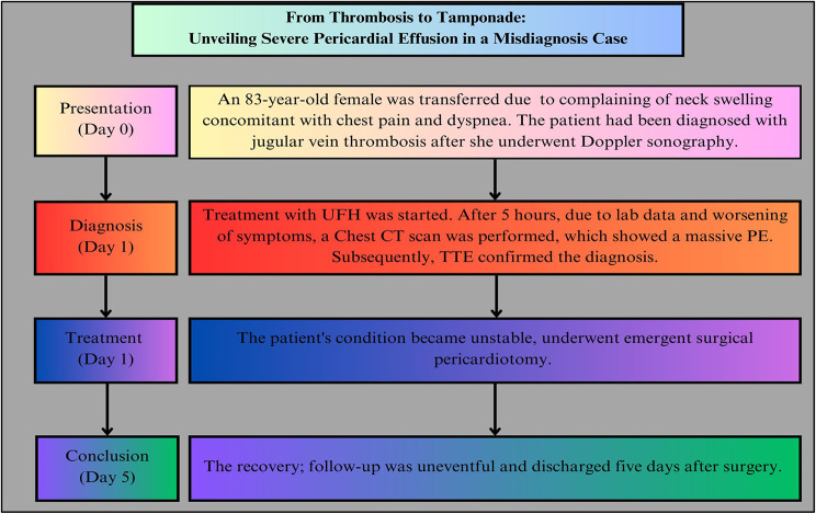
Background: Anticoagulants increase the risk of cardiac tamponade in patients with pericardial effusion (PE). Therefore, inappropriate administration of them in the presence of PE can lead to a catastrophic outcome. This study presents a patient with a provisional misdiagnosis of venous thromboembolism (VTE).
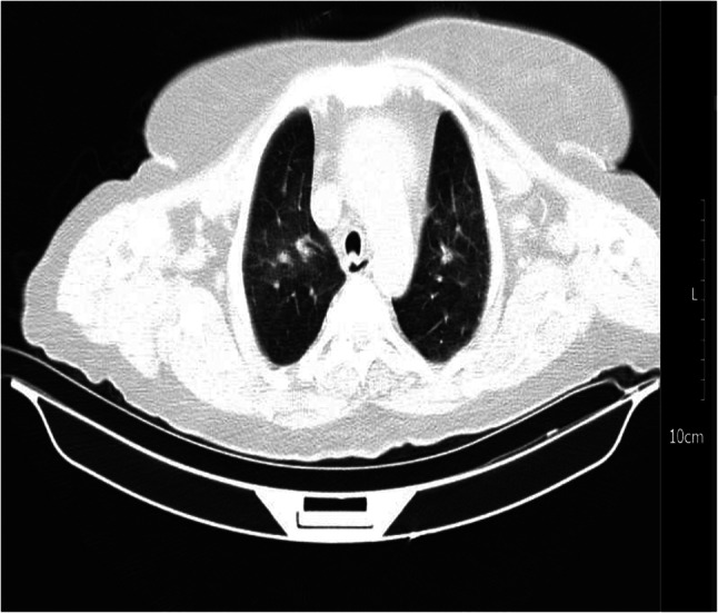
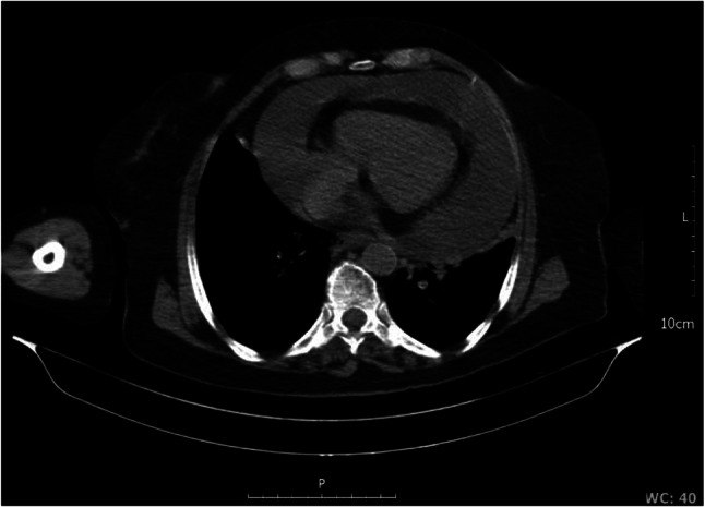
Case presentation: An 83-year-old Iranian female was transferred to the emergency department of a tertiary cardiology hospital complaining of neck swelling concomitant with chest pain and dyspnea. The patient had been diagnosed with jugular vein thrombosis in another local center, and since the chief complaint was neck swelling, she underwent Doppler sonography, and the diagnosis was confirmed. Subsequently, the treatment with unfractionated heparin was started. After 5 h, considering the worsening of symptoms with the suspicious diagnosis of COVID-19 based on her symptoms and laboratory data, a chest computed tomography scan was requested, which showed a massive PE. Subsequently, transthoracic echocardiography confirmed the diagnosis. The patient was immediately transferred to the operating room and underwent pericardiotomy. The post-surgery period was uneventful, and she was discharged 5 days later.
Conclusion: Patients with viral infections, specifically COVID-19, are at risk of undiagnosed severe pericardial effusions. Venous stasis in the jugular veins due to PE can mimic jugular vein thromboembolism, causing a wrong diagnosis. Since treating thrombosis can exacerbate tamponade to hemodynamic instability and collapse, sufficient investigation before starting anticoagulants is necessary.
Clinical key message: Distinguishing VTE from PE is not always straightforward. Therefore, it is important to ensure physicians have reached an appropriate level of certainty about their diagnosis by performing precise diagnostics before using anticoagulants. Mismanagement with anti-thrombotics can result in catastrophic consequences. Therefore, taking an accurate history, performing a precise physical examination, and using rapid and available diagnostic modalities can avoid delays in definitive management.
Keywords: Case report; Misdiagnosis; Pericardial effusion; Tamponade; Thromboembolism.
© 2024. The Author(s).
Conflict of interest statement
Declarations. Ethics approval: The study was performed in accordance with the ethical standards as laid down in the 1964 Declaration of Helsinki and its later amendments. Considering not including any revealed information about the patient and the breach of confidentiality, the committee waived the requirement for an ethics code. Consent to participate: Written informed consent was obtained from the patient. Consent for publication: Written informed consent was obtained from the patient for publication of this study and accompanying images. Competing interests: The authors declare no competing interests.
Figures


References
-
- Willner DA, Goyal A, Grigorova Y, Kiel J. Pericardial effusion. StatPearls. Treasure Island (FL): StatPearls Publishing Copyright © 2024. StatPearls Publishing LLC.; 2024. - PubMed
LinkOut - more resources
Full Text Sources
