. 2025 Mar;168(3):987-998.
doi: 10.1002/ijgo.16094. Epub 2025 Jan 4.

Complex cesarean section: Surgical approach to reduce the risks of intraoperative complications and postpartum hemorrhage

Collaborators, Affiliations

Complex cesarean section: Surgical approach to reduce the risks of intraoperative complications and postpartum hemorrhage

Albaro Jose Nieto-Calvache et al. Int J Gynaecol Obstet. 2025 Mar.

Abstract

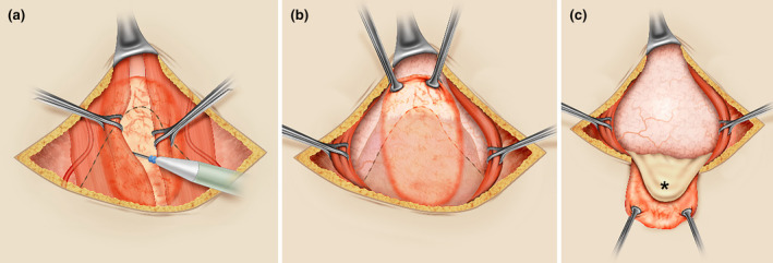
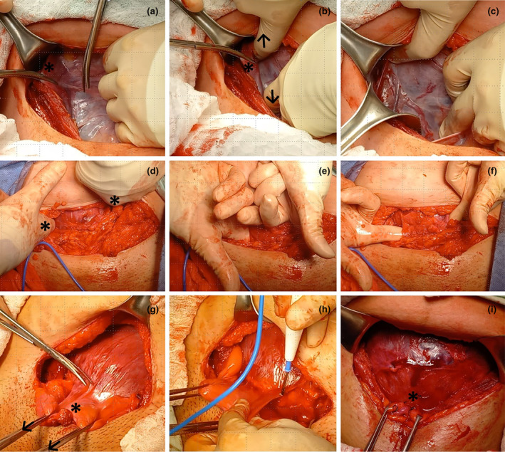
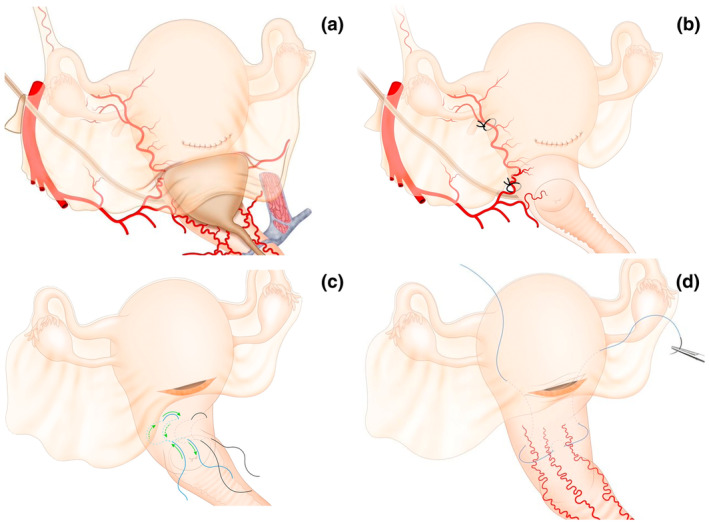
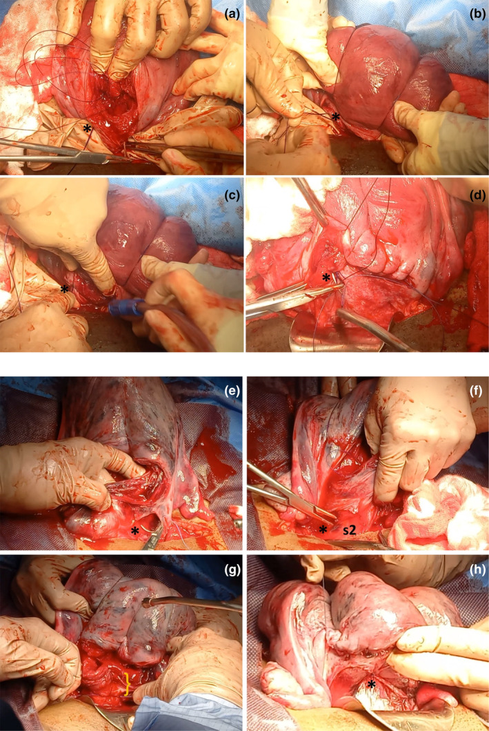
The incidence of cesarean section is dramatically increasing worldwide, whereas the training opportunities for obstetrician/gynecologists to manage complex cesarean section appear to be decreasing. This may be attributed to changing working hours directives and the increasing use of laparoscopy for gynecological surgical procedures, including in gynecological oncology. Various situations can create surgical difficulties during a cesarean section; however, two of the most frequent are complications from previous cesarean (myometrial defects, with or without placental intrusion and peritoneal adhesions) and the high risk of postpartum hemorrhage (uterine overdistension, abnormal placentation, uterine fibroids). Careful surgical dissection, with safe mobilization of the bladder and exposure of the anterior and lateral surfaces of the uterus, are pivotal steps for resolving the technical difficulties inherent in performing a complex cesarean section. We propose a standardized surgical protocol for women at risk of complex cesarean, including the antenatal identification of increased surgical risk, paramedian access to the pelvis, bladder dissection and mobilization, and the selection of a bleeding control strategy, considering uterine anatomy and the arterial pedicles involved in blood loss, which should be tailored to the individual case. We propose preoperative surgical planning to include consideration of the most common situations encountered during a complex cesarean, which facilitates anticipating an appropriate response for common possible scenarios, and can be adapted for low-, middle-, and high-resource settings. This protocol also highlights the importance of self-evaluation, continuous learning, and improvement activities within surgical teams.

Keywords: complex cesarean section; compressive uterine sutures; postpartum hemorrhage; protocolized treatment; surgical technique.


Conflict of interest statement

The authors declare no conflicts of interest.

Figures

FIGURE 1
FIGURE 1
Paramedian (preperitoneal) access to the pelvis. (a) In most cases, adhesions between the abdominal wall and intra‐abdominal structures occur along the midline (orange oval area), with the bladder being the most frequently adhered organ to the abdominal wall (interrupted black line). Using a transverse suprapubic skin incision, and after dissecting the abdominal fascia from the anterior surface of the rectus muscles, preperitoneal dissection is facilitated by retracting those muscles with Babcock forceps. The dissection begins at the medial edge of each muscle, moving laterally between the peritoneum and the muscle until reaching an area free of fibrosis. Attention should be paid to the inferior epigastric vessels on the lower lateral side of the rectus abdominis muscles when the dissection is extensive. (b) Upon reaching the lateral limit of the peritoneal adhesion area to the abdominal wall, the fibrotic area can be cut and mobilized (edges retracted with Allis forceps), also mobilizing the adhered bladder (interrupted black line) without risking injury. (c) After this type of dissection, the bladder (*) can be mobilized away from the anterior surface of the uterus, facilitating the hysterotomy.
FIGURE 2
FIGURE 2
Bladder mobilization during complex cesarean section. Exposure of the anterior surface of the uterine segment is fundamental for managing the surgical challenges in a complex cesarean section. (a–c) Opening of the parametrial space. Traction of the right round ligament (*) and incision of the anterior leaf of the broad ligament (a); digital opening (arrows) of the right parametrial space in a caudocephalic direction (b); mobilization of the round ligament with a retractor and exposure of the right lateral surface of the uterus (c). (d–f) Retrovesical bypass (Pelosi maneuver). The surgeon inserts their fingers (*) into both parametrial spaces (d) and moves them caudally to reach the medial paravesical spaces (e), then directs the fingertips toward the midline, at the level of the cervix, in the retrovesical space (f). (g‐i) Dissection of the retrovesical space. Facilitated by anterocaudal traction (45 degrees from the horizontal: Arrows) of the bladder (*) with Allis clamps (g), the peritoneum is incised at the vesicouterine fold. The lateral to medial approach, starting from the previously developed medial paravesical space (h), can be performed with scissors or energy, reducing the possibility of bladder injury or uterine laceration. Complete mobilization of the bladder (i) exposes the anterior surface of the uterine segment (*) and facilitates the selection and application of interventions such as low compression sutures (Ho Cho, B‐Lynch 2), uterine artery ligation, en‐bloc resection of lesions (leiomyoma, uterine dehiscence, placenta accreta spectrum) in that topography, or even hysterectomy.
FIGURE 3
FIGURE 3
Relationship between the mobilized and nonmobilized bladder with the performance of lower uterine compressive sutures and uterine artery ligation. The position of the bladder over the anterior surface of the lower uterine segment (a) makes it clear that controlling uterine bleeding from this area requires bladder mobilization. Ligation of the uterine artery on the lateral surface of the uterine segment requires that, in addition to the uterine vessels (artery and veins), a portion of the myometrium is included in the stitch to provide support for the suture (b). A deep stitch without bladder mobilization, even when applied from the posterior surface of the uterus, can lead to bladder tear, especially in patients with previous cesarean sections and adhesions between the uterus and bladder. Compression sutures for the lower uterine segment (c, d) are indicated in abnormal bleeding secondary to placenta previa. The lower part of the uterus receives its blood supply mainly from the colpouterine vessels that ascend from the vagina, and the most well‐known lower uterine compressive sutures are the Ho‐Cho suture (d) and the B‐Lynch 2 suture (e), whose application is impossible without prior bladder mobilization.
FIGURE 4
FIGURE 4
Importance of mobilization of the bladder to perform B‐Lynch suture. (a–d) Failed B‐Lynch in a patient with persistent bleeding after compression suturing and who was finally treated with hysterectomy. Application of the B‐Lynch suture without prior mobilization of the bladder implies that the initial (*) and final points of the suture are located very close to the lower edge of the hysterotomy (a) and that when compressing the uterus, the tension of the suture is supported by a very small portion of tissue (*) (b). This may cause the obstetrician to limit the force applied to the knot (*) due to the risk of lacerating the lower edge of the hysterotomy, resulting in a potentially suboptimal compressive effect (c). In these cases, the hysterorrhaphy may even pass just above the knot (*) of the compression suture (d). (e–h) Successful B‐Lynch. After dissection of the retrovesical space and exposure of the entire lower uterine segment, the first stitch of the compression suture (*) can be placed several centimeters caudal to the lower border of the hysterotomy (e). The same applies to the last stitch of the compression suture (*), which includes a significant amount of tissue in the lower part of the uterine segment in an area corresponding to sector 2 of uterine vascularization (s2), usually located behind the bladder (f). Including several centimeters of tissue at the lower border of the hysterotomy (}) allows the surgeon to exert greater compressive force with the suture without fear of tearing the tissue (g). Unlike in photo (d), in this case the knot of the compression suture (*) is located far from the hysterorrhaphy (h).
FIGURE 5
FIGURE 5
Broad ligament hematomas and hysterotomy tears. The most common bleeding sites in broad ligament hematomas are the lateral ends of the hysterotomy (due to persistent arterial bleeding from branches of the uterine artery) and the area where surgical sterilization is performed (due to bleeding from branches of the ovarian or uterine artery). During the exploration of hematomas, regardless of the size of the blood collection, the surgeon must specifically examine the suspected areas of bleeding origin (*) and for that it is essential to mobilize the bladder.
FIGURE 6
FIGURE 6
Uterine vascular pedicles. The uterus receives blood supply from three arterial pedicles. The most widely known are the ovarian (oA) and uterine arteries (uA), which supply the uterine body. The lower part of the uterus and the cervix receive arterial branches from the vagina, known as colpouterine arteries (cuA), typically identified as three main branches located in the midline (azygos vaginal artery–azA) and on either side of the anterior vaginal wall (*), reaching and anastomosing with branches of the uterine arteries. This last arterial pedicle (colpouterine arteries, branches off the inferior vaginal arteries–ivA) has been omitted in some anatomical descriptions, making it unknown to many obstetricians. Understanding these arterial pedicles allows for the establishment of two uterine vascular sectors. Sector S1 corresponding to the uterine body is primarily supplied by the uterine artery (uA) with a minor contribution from the ovarian artery (oA). Easy access to the vessels supplying this sector facilitates the control of bleeding from this part of the uterus. Conditions such as uterine atony can be successfully treated with multiple therapeutic options (uterine artery ligation, compression sutures, uterine artery embolization, etc.). Sector S2 corresponds to the lower uterine segment and cervix, receiving its main blood supply from the colpouterine arteries. Its subperitoneal and retrovesical location creates greater difficulty in controlling bleeding from this area (e.g. secondary to placenta previa or placenta accreta spectrum–PAS), necessitating bladder mobilization to perform procedures such as low compression sutures (B‐Lynch 2 or Ho‐Cho). The reference point separating sectors S1 and S2 is the peritoneal reflection (red dotted line), which is clearly visible during cesarean section when the bladder is empty. During prenatal ultrasonographic examination (or in magnetic resonance imaging) with a full bladder, the reference point is the midpoint of the posterior wall of the full bladder. In the female genital tract, a third vascular zone (sector S3), corresponds to the vagina, supplied by vaginal arteries, branches off the internal pudendal artery (originating from the posterior division of the internal iliac artery). Controlling bleeding from this zone (e.g. in complex vaginal tears associated with compound fetal presentations or instrumental vaginal delivery) demands exposure of the external surface of the vagina by dissecting the prevesical space (Retzius).
FIGURE 7
FIGURE 7
Immediate temporary control of bleeding during cesarean section. Regardless of the cause of uterine or pelvic bleeding, there are options for immediate and temporary hemorrhage control (primary hemostasis). In cases of uterine bleeding, the tourniquet with a sterile latex glove (a) applied below the level of bleeding (b) allows for bleeding cessation while other definitive control strategies are applied (secondary hemostasis). Manual compression of the infrarenal aorta (zone 3 of the aorta) provides immediate control of pelvic bleeding of any etiology (primary hemostasis). A healthcare professional positions themselves beside the patient and, after locating the aorta by palpation, compresses it against the lumbar vertebrae with the weight of their torso, positioning their arm at a 90° angle to the horizontal (c), with the elbow fully extended. A step stool should be used to achieve this position and maintain it as long as necessary (up to 60 continuous minutes without significant metabolic impact) so that surgeons can apply other definitive bleeding control interventions (secondary hemostasis).

Similar articles

References

    1. Betran AP, Ye J, Moller AB, Souza JP, Zhang J. Trends and projections of caesarean section rates: global and regional estimates. BMJ Glob Health. 2021;6:e005671. - PMC - PubMed
    1. Silver RM, Landon MB, Rouse DJ, et al. Maternal morbidity associated with multiple repeat cesarean deliveries. Obstet Gynecol. 2006;107:1226‐1232. - PubMed
    1. Jauniaux E, Fox KA, Einerson B, Hussein AM, Hecht JL, Silver RM. Perinatal assessment of complex cesarean delivery: beyond placenta accreta spectrum. Am J Obstet Gynecol. 2023;229:129‐139. - PubMed
    1. Futterman ID, Conroy EM, Chudnoff S, Alagkiozidis I, Minkoff H. Complex obstetrical surgery: building a team and defining roles. Am J Obstet Gynecol MFM. 2024;6:101421. - PubMed
    1. Gialdini C, Chamillard M, Diaz V, et al. Evidence‐based surgical procedures to optimize caesarean outcomes: an overview of systematic reviews. EClinicalMedicine. 2024;72:102632. - PMC - PubMed

MeSH terms