. 2025 Feb;603(4):971-987.
doi: 10.1113/JP286685. Epub 2025 Jan 4.

Enhanced myocardial perfusion in late gestation fetal lambs with impaired left ventricular inflow

Affiliations

Enhanced myocardial perfusion in late gestation fetal lambs with impaired left ventricular inflow

Matthew W Hagen et al. J Physiol. 2025 Feb.

Abstract

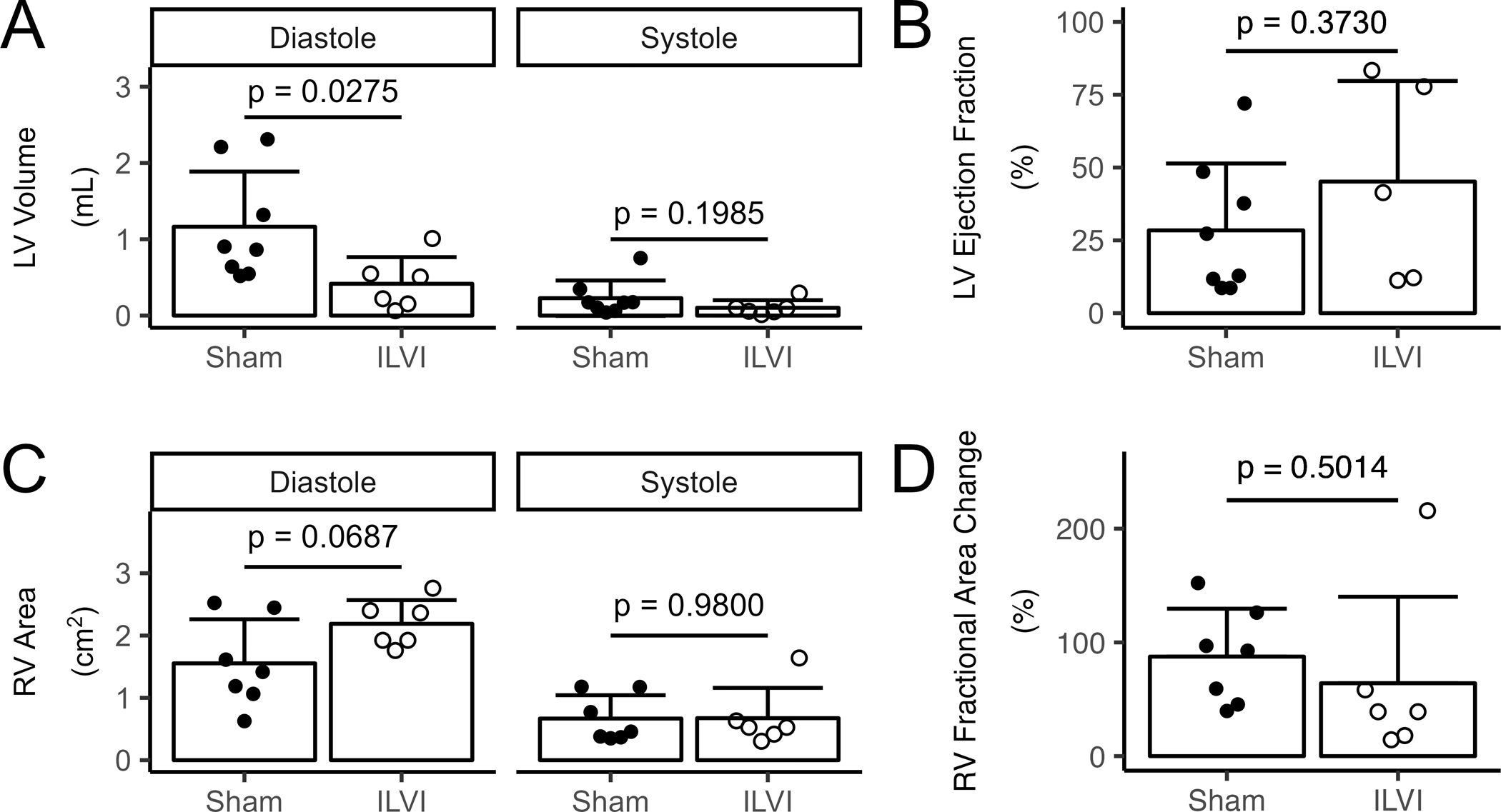
Robust preclinical models of asymmetric ventricular loading in late gestation reflecting conditions such as hypoplastic left heart syndrome are lacking. We characterized the morphometry and microvascular function of the hypoplastic left ventricle (LV) and remaining right ventricle (RV) in a sham-controlled late gestation fetal lamb model of impaired left ventricular inflow (ILVI). Singleton fetuses were instrumented at ∼120 days gestational age (dGA; term is ∼147 days) with vascular catheters, an aortic flow probe and a deflated left atrial balloon. Balloons in ILVI fetuses were inflated over the 8 day study until aortic output was eliminated; Sham balloons remained deflated. At the study end-point (∼134 dGA), cardiac function was assessed by echocardiography, microvascular perfusion of each free wall was measured by myocardial contrast echocardiography (MCE) and terminal morphometric data were collected. During the chronic study, flow through the ascending aorta of ILVI fetuses fell from 389 to -48 mL min-1 with minimal changes to other haemodynamics or blood chemistry. End-point echocardiography and morphometry similarly showed significant and meaningful reductions in ILVI LV chamber volume and wall mass without statistically significant changes in RV size relative to Shams. MCE revealed modestly increased LV perfusion and profoundly increased RV perfusion in ILVI fetuses. Our model displays effective LV hypoplasia with preserved overall fetal health, and our finding of increased RV myocardial perfusion may indicate active vascular remodelling in response to the experimental lesion. KEY POINTS: Hypoplastic left heart syndrome can be caused by insufficient inflow of blood to the fetal left ventricle. We found that eliminating fetal left ventricular input for 8 days reduced left ventricular size and volume, with minimal effects on the right ventricle or overall fetal health. Blood and oxygen delivery increased significantly in the right ventricle and slightly in the hypoplastic left ventricle. Our results suggest functional and anatomical adaptation of the fetal coronary microvasculature to univentricular right heart conditions.

Keywords: coronary microvascular function; development; fetus; heart; hypoplastic left heart; vascular remodelling.


Conflict of interest statement

The authors declare no competing interests.

Figures

Figure 1.
Figure 1.. Hemodynamics during chronic study
(A) Representative ascending aortic flow traces from sham (left) and ILVI (right) fetuses recorded at study day 0 (dotted lines) and study day 8 (solid lines) at 200 Hz showing two consecutive cardiac cycles. (B) Flow through the ascending aorta was affected by study intervention (interaction p = 7.4×10⁻⁹). Šídák contrasts: ILVI aortic flow was lower at day 8 than day 0 (p = 7.0×10⁻¹⁰), Sham and ILVI were significantly different at day 4 (p = 0.0010) and day 8 (p = 0.0001). (C) Aortic pressure was not significantly affected by study intervention (time p = 0.3910, group p = 0.1581, interaction p = 0.1455). (D) Right atrial (RA) pressure showed a tendency toward a difference between groups (p = 0.0519) and no significant differences by time (p = 0.9583) and no significant interaction (p = 0.0983). (E) Heart rate was significantly affected by time (p = 1.3×10⁻⁷), however there were no significant group (p = 0.1134) or interaction effect (p = 0.1120). Shams (n = 7 in B and 9 in C-E) illustrated by closed circles and solid black lines, ILVI (n = 6 in B and 7 in C-E) by open magenta circles and dotted magenta lines in panels B-D (refer to legend in C). Data are means ± SD; each dot represents one animal at one timepoint. Data analyzed with linear mixed effects. Post-hoc Šídák contrasts: between-group **p < 0.01, *** p < 0.001, within-group ††† p < 0.001.
Figure 2.
Figure 2.. Circulating factors
(A) Log high sensitivity Troponin I concentration was affected by study intervention (p = 0.0014). Šídák contrasts: ILVI day 8 was significantly higher than ILVI day 0 (p = 0.0001) and greater than Sham day 8 (p = 0.001). (B) No significant changes in atrial natriuretic peptide concentration were seen (group p = 0.0802, time p = 0.4119, interaction p = 0.8768). (C) Plasma renin activity was not affected by the study intervention (group p = 0.4137, time p = 0.0759, interaction p = 0.2546). Shams (n = 9) illustrated by closed circles and solid black lines, ILVI (n = 7) by open circles and dotted magenta lines. Data are means ± SD; each dot represents one animal at one timepoint. Data analyzed with linear mixed effects. Šídák contrasts: between-group **p < 0.01, within-group ††† p < 0.001.
Figure 3.
Figure 3.. Chamber morphometry and systolic function measured by echocardiography
(A) Left ventricular (LV) volume was significantly greater in Sham than ILVI fetuses at end diastole but not different at end systole; Sham (n = 8), ILVI (n = 6). (B) LV ejection fraction was not significantly different between groups; Sham (n = 8), ILVI (n = 5). (C) Right ventricular (RV) area tended to be higher in ILVI relative to Sham fetuses but did not reach statistical significance; Sham (n = 7), ILVI (n = 6). (D) RV fractional area change (long axis) was not different between groups; Sham (n = 7), ILVI (n = 6). Columns are means and error bars are + SD, each dot represents one animal, all comparisons by t-test.
Figure 4.
Figure 4.. Myocardial perfusion by myocardial contrast echocardiography
(A) Microvascular blood volume (MBV) was significantly greater in ILVI fetuses than Shams in both RV and LV. (B) Flux rate was faster in ILVI RV than Shams, and tended to be faster in ILVI LV than Shams. (C) Microvascular blood flow (MBF) was higher in ILVI than Sham left ventricular (LV) and right ventricular (RV) free walls. (D) Oxygen delivery was not different in LV but was greater in ILVI than Sham RV. Columns are means and error bars are + SD; each dot represents one animal; nonparametrically distributed data (RV flux rate) analyzed by Mann-Whitney U-test, all others by t-test; LV: Sham (n = 6), ILVI (n = 5); RV: Sham (n = 5), ILVI (n = 5).
Figure 5.
Figure 5.. Myocardial perfusion by MCE during adenosine-mediated hyperemia
(A) Microvascular blood volume (MBV) during hyperemia was not different in LV or RV free walls. (B) Flux rate during hyperemia tended to be faster in ILVI than Sham RV free walls and was significantly faster in ILVI than Sham RV free walls. (C) Microvascular blood flow (MBF) during adenosine-mediated hyperemia was higher in ILVI than Sham left ventricular (LV) and right ventricular (RV) free walls. Columns are means and error bars are + SD; each dot represents one animal; nonparametrically distributed data (all microvascular flow, RV flux rate) analyzed by Mann-Whitney U-test, all others by t-test; LV: Sham (n = 6), ILVI (n = 5); RV: Sham (n = 5), ILVI (n = 5).
Figure 6.
Figure 6.. Postmortem cardiac morphometry
Two representative photographs each of (A) Sham and (B) ILVI perfusion-fixed hearts. Hearts were photographed anterior face-up on a mat with a 1 cm × 1cm grid. White arrows identify the left ventricle (LV). (C) There was no significant difference in whole heart weights. (D) ILVI left and right atria were larger than Sham. (E) ILVI LV free walls weighed less than Sham; there was no difference in right ventricular (RV) free wall or ventricular septal weights. Columns are means and error bars are + SD, each dot represents one animal; data analyzed with t-test; Sham (n = 8), ILVI (n = 7).

References

    1. Alanne L, Bhide A, Lantto J, Huhta H, Kokki M, Haapsamo M, Acharya G & Räsänen J (2021). Nifedipine disturbs fetal cardiac function during hypoxemia in a chronic sheep model at near term gestation. American Journal of Obstetrics and Gynecology 225, 544.e1–544.e9. - PubMed
    1. Bader D, Kugelman A, Lanir A, Tamir A, Mula E & Riskin A (2006). Cardiac troponin I serum concentrations in newborns: A study and review of the literature. Clinica Chimica Acta 371, 61–65. - PubMed
    1. Barbera A, Giraud GD, Reller MD, Maylie J, Morton MJ & Thornburg KL (2000). Right ventricular systolic pressure load alters myocyte maturation in fetal sheep. Am J Physiol Regul Integr Comp Physiol 279, R1157–1164. - PubMed
    1. Broberg CS, Giraud GD, Schultz JM, Thornburg KL, Hohimer AR & Davis LE (2003). Fetal anemia leads to augmented contractile response to hypoxic stress in adulthood. Am J Physiol Regul Integr Comp Physiol 285, R649–655. - PubMed
    1. Brooks PA, Khoo NS, Mackie AS & Hornberger LK (2012). Right Ventricular Function in Fetal Hypoplastic Left Heart Syndrome. Journal of the American Society of Echocardiography 25, 1068–1074. - PubMed

LinkOut - more resources