

Trigeminal artery anatomical aspects
- PMID: 39754664
- PMCID: PMC11700043
- DOI: 10.1007/s00276-024-03553-0
Trigeminal artery anatomical aspects
Abstract
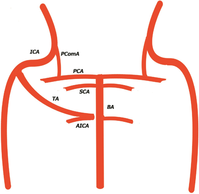
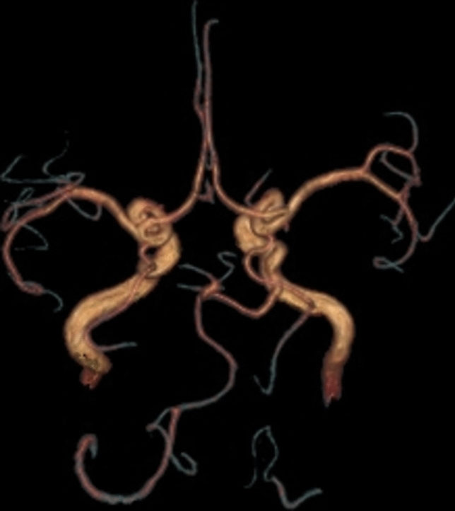
Purpose and background: The trigeminal artery is a rare anatomical variant, representing an embryonic vestige of the anastomosis between the internal carotid artery and the posterior circulator system, that can be asymptomatic or could have vast clinical manifestations produced by insufficient flow or by vascular nervous conflicts. This study is an anatomical presentation of 3 trigeminal artery cases observed at Medimar Imagistic Services Constanta.
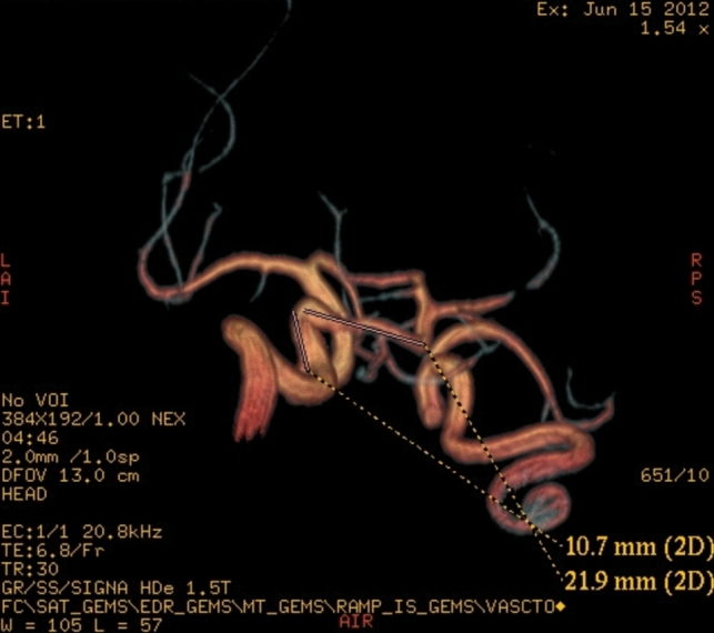
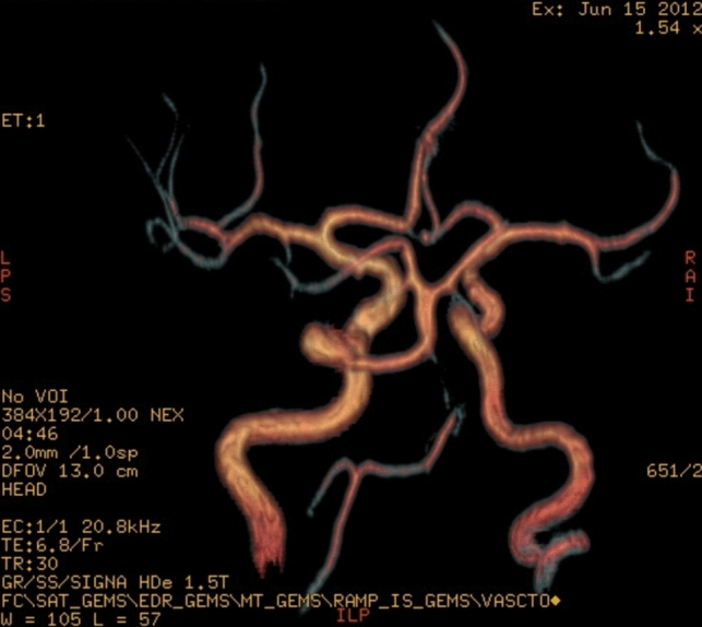
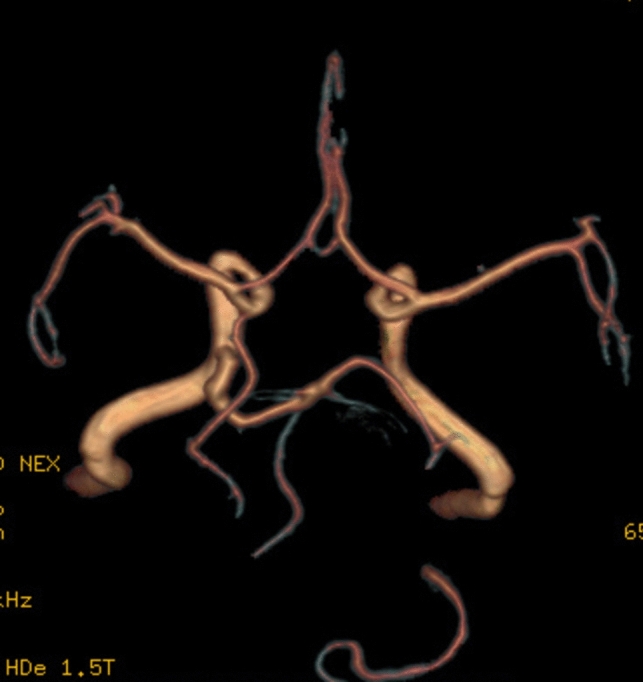
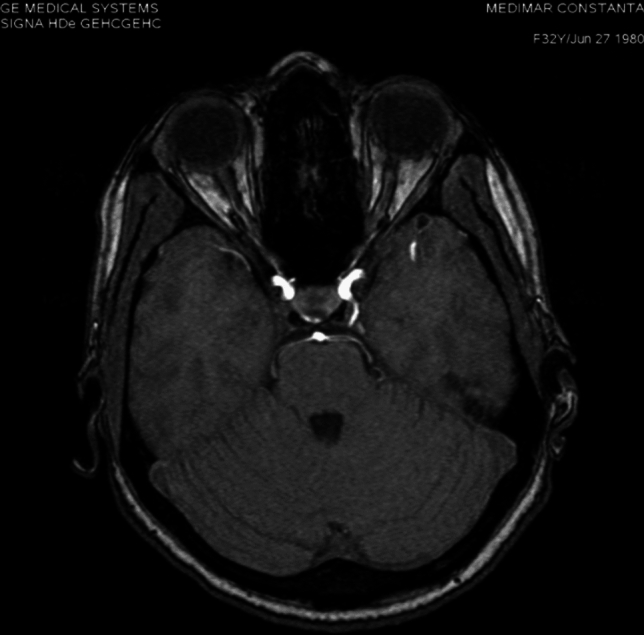
Methods: The 3 trigeminal artery cases were discovered on a 860 magnetic resonance angiographies (0.35% of cases), made on a GE HD/e 8ch 1.5 T.
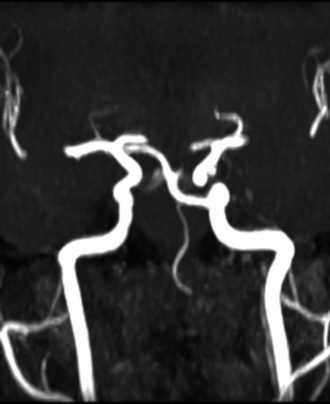
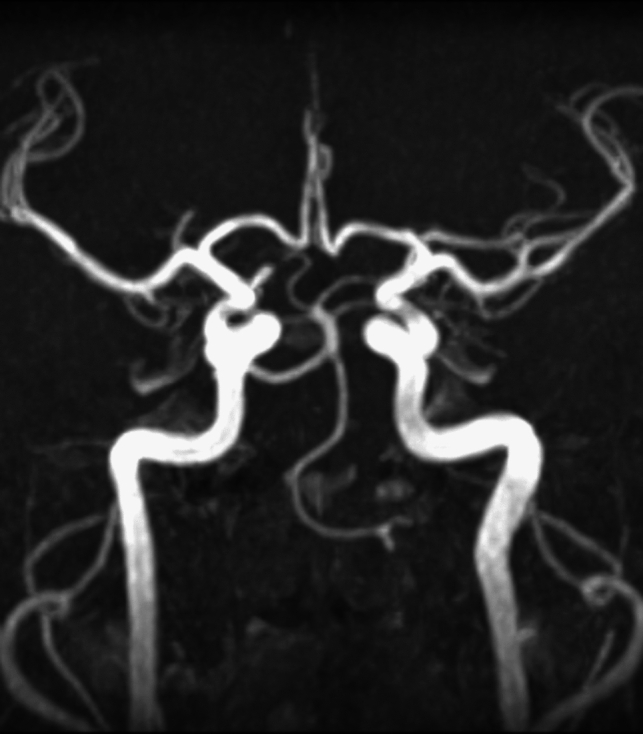
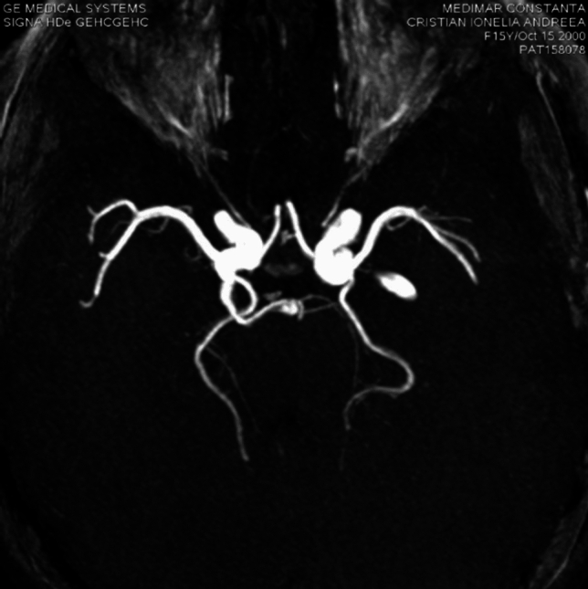
Results: In all 3 cases, the arteries were rising from the right internal carotid artery and one from the left internal carotid artery, in 2 cases the origin was on the superior surface, and in one case, on the anterolateral surface, in all 3 cases from C4 horizontal cavernous segment. The artery caliber was between 2.7 mm and 5.1 mm; the artery length was between 26 and 32 mm. Other associated vascular malformations were: partial or total basilar artery hypoplasia, in one case posterior communicant artery agenesis, contralateral vertebral artery hypoplasia, posterior cerebral artery hypoplasia (in 2 cases), and no anastomosis between the P1 segment of the posterior cerebral artery with the posterior communicating artery (one case).
Conclusions: While the trigeminal artery is a rare anatomical variant, it's still a very important vessel situated in the posterior cerebral fossa, which needs to be taken into account in the case of neurosurgical interventions.
Keywords: Anatomic aspects; Trigeminal artery.
© 2025. The Author(s).
Conflict of interest statement
Declarations. Conflict of interests: The authors declare no competing interests. Ethical Approval: The study is in accordance with the local Ethics Commission (34490 / 08.08.2019) within the institution and respects the principles of the General Regulation on Personal Data Protection (EU) 679/2016. The research was conducted in accordance with the Helsinki Declaration and in compliance with all relevant national regulations regarding patient studies. Informed consent was obtained from the patients.
Figures


References
-
- Allen JW, Alastra AJ, Nelson PK (2005) Proximal intracranial internal carotid artery branches: prevalence and importance for balloon occlusion test. J Neurosurg. 10.3171/jns.2005.102.1.0045 - PubMed
-
- Battista RA, Kwartler JA, Martinez DM (1997) Persistent trigeminal artery as a cause of dizziness. Ear Nose Throat J 76:43–45. 10.1177/014556139707600112 - PubMed
-
- Chen YC, Li MH, Chen SW, Hu DJ, Qiao RH (2011) Incidental findings of persistent primitive trigeminal artery on 3-dimensional time-of-flight magnetic resonance angiography at 3.0 T: An analysis of 25 cases. J Neuroimaging 21:152–158. 10.1111/j.1552-6569.2010.00472.x - PubMed
Publication types
MeSH terms
LinkOut - more resources
Full Text Sources
Miscellaneous
