. 2025 Jan 4;15(1):803.
doi: 10.1038/s41598-024-82331-9.

Oncolytic alphavirus-induced extracellular vesicles counteract the immunosuppressive effect of melanoma-derived extracellular vesicles

Affiliations

Oncolytic alphavirus-induced extracellular vesicles counteract the immunosuppressive effect of melanoma-derived extracellular vesicles

Darshak K Bhatt et al. Sci Rep. .

Abstract

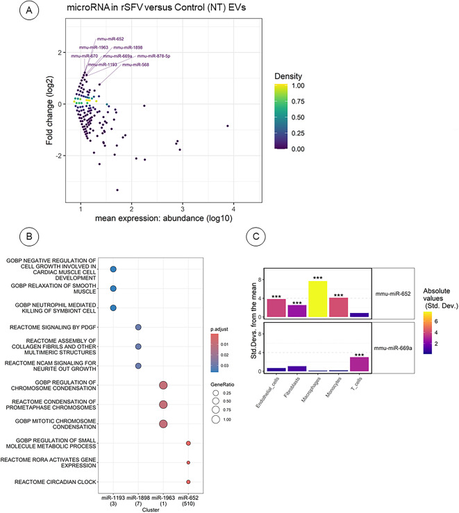
Extracellular vesicles (EVs)-mediated communication by cancer cells contributes towards the pro-tumoral reprogramming of the tumor microenvironment. Viral infection has been observed to alter the biogenesis and cargo of EVs secreted from host cells in the context of infectious biology. However, the impact of oncolytic viruses on the cargo and function of EVs released by cancer cells remains unknown. Here we show that upon oncolytic virotherapy with Semliki Forest virus-based replicon particles (rSFV), metastatic melanoma cells release EVs with a distinct biochemical profile and do not lead to suppression of immune cells. Specifically, we demonstrate that viral infection causes a differential loading of regulatory microRNAs (miRNAs) in EVs in addition to changes in their physical features. EVs derived from cancer cells potentially suppress splenocyte proliferation and induce regulatory macrophages. In contrast, EVs obtained from rSFV-infected cells did not exhibit such effects. Our results thus show that rSFV infection induces changes in the immunomodulatory properties of melanoma EVs, which may contribute to enhancing the therapeutic efficacy of virotherapy. Finally, our results show that the use of an oncolytic virus capable of a single-round of infection allows the analysis of EVs secreted from infected cells while preventing interference from extracellular virus particles.

Keywords: Extracellular vesicles; Immunomodulatory; Melanoma; Oncolytic virus.


Conflict of interest statement

Declarations. Competing interests: The authors declare no competing interests.

Figures

Fig. 1
Fig. 1
Isolation of tumor-derived EVs upon exposure to rSFV replicon particles. (A) Semliki Forest virus replicon particles were engineered by replacing the structural genes in the genome with a GFP-transgene (left) to enable a single round of infection (right). (B) Illustration of the experimental scheme for rSFV infection of B16F10 cells followed by a wash 2 h post-infection to remove extracellular virus particles and incubation for 24 h before the collection of supernatants followed by EV isolation through sequential ultracentrifugation steps. (C) Absolute vesicle yield from infected (rSFV EVs) or non-infected B16F10 cells (NT EVs) in different batches. (D) Decrease in the number of viable B16F10 cells upon rSFV infection. (E) Vesicles per cell count based on number of cells present at the time of EV isolation. (F) Size distribution of EVs from infected and non-infected cells. In (CE) n = 3 independent batches of EV-preparation are shown. Data are presented as mean values. One-way ANOVA followed by Bonferroni’s multiple comparisons test was performed.
Fig. 2
Fig. 2
Characterization of tumor-derived EVs upon exposure to rSFV replicon particles. (A) Zeta potential of SFV EVs and NT EVs. (B) Western blot image for validation of expression of CD63 and CD9 as EV membrane markers in the EV-preparations. (C) Transmission electron microscopy of vesicles from non-infected and SFV-infected melanoma cells. In (A) Data are presented as mean values.
Fig. 3
Fig. 3
The effect of rSFV-induced EVs on tumor establishment. (A) Overall scheme of the experimental steps where B16F10 cells were pre-treated with EVs from infected (SFV EVs) or non-infected cells (NT EVs) daily for 5 days and then collected for an in vivo tumor engraftment assay or an in vitro clonogenic assay. (B) Percentage of animals free from tumor and (C) related tumor growth rate upon injection with B16F10 cells pretreated with SFV EVs or NT EVs (n = 5 for each group). (D) Frequency of colonies formed from 200 cells plated after 7 days of clonogenic assay. (E) Representative images of colonies in a well formed by B16F10 cells from different conditions. In (D,E) n = 12 replicates from 4 independent experiments. One-way ANOVA followed by Bonferroni’s multiple comparisons test was performed. Data are presented as mean values ± SD.
Fig. 4
Fig. 4
Immunoregulatory phenotype of melanoma cells upon exposure to EVs from rSFV-infected or non-infected cells. (A) Experimental scheme to study changes in gene expression of melanoma cells upon vesicle treatment. (B) Heatmap of gene expression upon treatment with vesicles from non-infected or SFV-infected melanoma cells. (C) Experimental setup of co-culture of vesicle treated melanoma cells and splenocytes. (D) Flow cytometry analysis of tumor cell-death and CD4 and CD8 T cell activation. Frequency of active (E) cytotoxic CD8 T cells or (F) helper CD4 T cells expressing CD69. (G) Frequency of melanoma cells that are dead, apoptotic (Casp3/7+) or non-apoptotic (Casp3/7-). In (A) n = 4 replicates. In (B) 2−ΔΔCt values were calculated with respect to endogeneous (Hprt) gene expression and plotted as fold change data points on a log2 axis for 3 replicates. Cells not treated with EVs (No EVs group) was used as reference sample. In (C) the replicates represent data from 2 splenocytes isolated from healthy mice. In E, F and G, the labels A and B represent data from the two different splenocytes.
Fig. 5
Fig. 5
The effect of rSFV-induced EVs on macrophage polarization. (A) Illustration of the experiment where bone-marrow derived macrophages were polarized to inflammatory M1-like or regulatory M2-like macrophages in presence of EVs from infected (SFV EVs) or non-infected cells (NT EVs). Polarization towards an M1-like phenotype was done through treatment with LPS and IFN-y, while towards an M2-like phenotype was done with IL-4. Macrophage polarization was assessed by quantification of mRNA expression of (B) Mrc1, (C) Arg1, (D) Il12, (E) iNos, and (F) Il10, with Hprt used as an endogenous housekeeping gene. In (BF) bone-marrow derived macrophages tested from n = 3 animals in independent experiments. Data are presented as mean values ± SD.
Fig. 6
Fig. 6
The effect of rSFV-induced EVs on splenocyte activation and proliferation. (A) Illustration of the experimental steps where freshly isolated splenocytes from mice were labeled with CFSE and then activated with PMA and ionomycin in the presence of EVs from infected (SFV EVs) or non-infected cells (NT EVs). 48 h post treatment, CFSE-labeled splenocytes were (B) counted manually and (C) analyzed by flow cytometry to observe CFSE-based proliferation profiles. Splenocytes were isolated from n = 4 animals for independent experiments. Data are presented as mean values ± SD.
Fig. 7
Fig. 7
Differentially Expressed Vesicular miRNAs in rSFV EVs. (A) Scatter plot illustrating vesicular miRNAs with elevated fold changes in EVs derived from rSFV cells. Labeled miRNAs highlight key regions based on fold change and mean expression levels (abundance). The color scale represents the density of miRNA probes overlapping the same position. (B) Dot plot depicting enriched pathways (adjusted p < 0.05) identified through hallmark (H), curated (C2), and ontology (C5) gene sets from the Molecular Signature Database (MSigDB). (C) Immune cell types with enriched expression of miRNA targets, based on data from the Immunologic Genome Project (ImmGen). *** indicates adjusted p < 0.001.

Similar articles

Cited by

References

    1. Cocozza, F., Grisard, E., Martin-Jaular, L., Mathieu, M. & Théry, C. SnapShot: extracellular vesicles. Cell182, 262–262e1. 10.1016/j.cell.2020.04.054 (2020). - PubMed
    1. Colombo, M., Raposo, G. & Théry, C. Biogenesis, secretion, and intercellular interactions of exosomes and other extracellular vesicles. Annu. Rev. Cell Dev. Biol.30, 255–289. 10.1146/annurev-cellbio-101512-122326 (2014). - PubMed
    1. Pegtel, D. M. & Gould, S. J. Exosomes Annu. Rev. Biochem.88, 487–514. 10.1146/annurev-biochem-013118-111902. (2019). - PubMed
    1. Schorey, J. S., Cheng, Y., Singh, P. P. & Smith, V. L. Exosomes and other extracellular vesicles in host-pathogen interactions. EMBO Rep.16, 24–43. 10.15252/embr.201439363 (2015). - PMC - PubMed
    1. Alenquer, M. & Amorim, M. Exosome biogenesis, regulation, and function in viral infection. Viruses7, 5066–5083. 10.3390/v7092862 (2015). - PMC - PubMed

Publication types

LinkOut - more resources