Review
. 2025 May;45(3):968-984.
doi: 10.1002/med.22099. Epub 2025 Jan 5.

Spatiotemporal Control Over Circadian Rhythms With Light

Affiliations
Review

Spatiotemporal Control Over Circadian Rhythms With Light

Dušan Kolarski et al. Med Res Rev. 2025 May.

Abstract

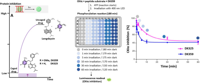
Circadian rhythms are endogenous biological oscillators that synchronize internal physiological processes and behaviors with external environmental changes, sustaining homeostasis and health. Disruption of circadian rhythms leads to numerous diseases, including cardiovascular and metabolic diseases, cancer, diabetes, and neurological disorders. Despite the potential to restore healthy rhythms in the organism, pharmacological chronotherapy lacks spatial and temporal resolution. Addressing this challenge, chrono-photopharmacology, the approach that employs small molecules with light-controlled activity, enables the modulation of circadian rhythms when and where needed. Two approaches-relying on irreversible and reversible drug activation-have been proposed for this purpose. These methodologies are based on photoremovable protecting groups and photoswitches, respectively. Designing photoresponsive bioactive molecules requires meticulous structural optimization to obtain the desired chemical and photophysical properties, and the design principles, detailed guidelines and challenges are summarized here. In this review, we also analyze all the known circadian modulators responsive to light and dissect the rationale following their construction and application to control circadian biology from the protein level to living organisms. Finally, we present the strength of a reversible approach in allowing the modulation of the circadian period and the phase.

Keywords: azobenzene; circadian clock; circadian rhythm; light; photopharmacology; photoswitch; photo‐removable protecting group.


Conflict of interest statement

The authors declare no conflicts of interest.

Figures

Figure 1
Figure 1
Origin and hierarchical organization of circadian rhythms. Parts of the figure were downloaded from www.freepik.com and modified accordingly.
Figure 2
Figure 2
Disruption of a peripheral circadian clock and two approaches to restoring healthy rhythms: pharmacological (systemic action) and chrono‐photopharmacological (spatiotemporal action). Parts of the figure were downloaded from www.freepik.com and modified accordingly.
Figure 3
Figure 3
The endogenous regulation of circadian rhythms in mammals and an outline of small light‐responsive molecules that can be used for its reversible modulation. (A) Circadian regulation on the molecular level consisted of the primary and secondary circadian loop as well as posttranslational modification. Genes are denoted in italics, while promotors, proteins, and enzymes are in capital letters. (B) Photoresponsive modulators of the circadian rhythm, color‐coded based on the color of light they respond to for activation.
Figure 4
Figure 4
Principle of reversible photopharmacology. Adapted and modified with permission from Ref. [88] Copyright © 2017 American Chemical Society.
Figure 5
Figure 5
Scheme of (A) irreversible and (B, C) reversible approach to circadian rhythm modulation. The uncaging and trans‐to‐cis isomerization are indicated with a purple bulb, cis‐to‐trans isomerization using a yellow light bulb, and the thermal back‐isomerization in orange.
Figure 6
Figure 6
Photoresponsive modulators (violet, green, and blue) of the circadian rhythm and their parent structures (gray). Parts of the modulators used for creating the photoresponsive modulators are shown in orange, while the colors of the photoresponsive modulators refer to the wavelength of light they respond to.
Figure 7
Figure 7
Irreversible approach to CKIα activity control: scheme of the approach and the experimental setup, as well as response curves of CKIα inhibition and irradiation time. Part of the figure was adapted with permission from Ref [80] Copyright © 2019 American Chemical Society.
Figure 8
Figure 8
Control of CKI (CKIα and CKIẟ) and RORγ activity using photoswitches. (A) Schematic presentation of two possible enzyme activity modulation approaches using photoswitches – photoactivation and photodeactivation. (B, C) Two types of photoswitches based on Longdaysin inhibitor, their chemical properties, and protein inhibitions in the dark and upon light irradiation. (D) Light‐responsive CKIẟ inhibitors based on LH846. (E) Photoswitchable modulators of RORγ activity. Parts of the figure were adapted with permission from Ref [78, 79, 81, 83]. Copyright © 2021 The Royal Society of Chemistry, © 2021 Springer Nature, © 2022 MDPI, and © 2024 Wiley.
Figure 9
Figure 9
Irreversible control of the circadian period lengthening in cells. Modulating the circadian period in cells by precisely controlled irradiation (A) in the presence of caged Longdaysin modulators DK325 and DK359. (B) Luminescence rhythms and the quantified period change of the U2OS cells treated with the caged modulators followed by dark conditions (0 min) or upon irradiation with violet light (30 min). (C) Circadian rhythms displaying the effect of violet light irradiation on Day 3 after the caged modulator was applied, and the corresponding period change during the first and second halves of the experiment. Parts of the figure were adapted with permission from Ref [80] Copyright © 2019 American Chemical Society.
Figure 10
Figure 10
Reversible control of the circadian period lengthening in cells. (A) Photo‐ and thermal‐isomerization of compound 9trans, a potent modulator, and a weak cis. (B) Concentration‐ and light‐dependent period lengthening measured in U2OS cells. The modulator was applied to the cells and kept continuously in the dark (black), irradiated with green light to reach the corresponding PSD (green), irradiated with green followed by violet light (violet), and irradiated with violet light (gray). (C, D) The period change in a long‐term experiment where (C) the modulator was deactivated by green light on Day 3 or (D) deactivated with green light upon addition to the cells followed by reactivation with violet light on Day 3. Parts of the figure were adapted with permission from Ref [78] Copyright © 2021 Springer Nature.
Figure 11
Figure 11
A reversible modulator of the circadian period based on the CRY1 selective binder, TH129. (A) Azologization of TH129 and photoisomerization of GO1423; (B) A rationale behind azologization of benzophenones; (C) The analysis of data from Cambridge Structural Database (CSD) and comparison between distributions of the ring angles and distances of benzophenones, cis‐ and trans‐azobenzenes; (D) Period lengthening of transGO1423 (“dark,” “green + violet,” and “dark + violet”) and cisGO1423 (“green”) as well as the period lengthening of TH129 under the same irradiation conditions. Parts of the figure were adapted with permission from Ref. [82] Copyright © 2021 American Chemical Society.
Figure 12
Figure 12
Photodosing of the circadian period in tissues. (A) Luminescence rhythms of the spleen explants in the presence of caged modulators (DK325 and DK359) kept in the dark (0 min) or irradiated with violet light (30 min). (B) Quantified period lengthening of both modulators and Longdaysin in the dark and upon irradiation at two concentrations (8 and 24 µM). (C) The period change correlated with violet light exposure at a single concentration (24 µM, 0–30 min irradiation). (D) Luminescence rhythms and quantified period change in the SCN tissue explants. DK359 was applied at the beginning of the assay and irradiated with violet light (5 min) on Day 5, whereas the other half was continuously kept in the dark. The figure was adapted with permission from Ref [80] Copyright © 2019 American Chemical Society.
Figure 13
Figure 13
Reversible modulation of the circadian period in tissue. (A–C) The peripheral tissue (spleen) explants were treated with trans9 and kept in the dark (black line) or irradiated with a green light at the beginning of the assay (green line). (D) SCN luminescence rhythms and the corresponding period changes. Trans9 was added on Day 4, and the explants were irradiated with green light on Day 8 to suppress period lengthening. The figure was adapted with permission from Ref. [78] Copyright © 2021 Nature Springer.
Figure 14
Figure 14
Modulation of circadian rhythm in zebrafish larvae. (A) Luminescence rhythms of larvae treated with DMSO or DK359 followed by the dark or irradiated for 10 min with violet light. (B) Period change in larvae depending on the irradiation time. The figure was adapted with permission from Ref [80] Copyright © 2019 American Chemical Society.
Figure 15
Figure 15
Circadian phase modulation with compound 9. (A, B) Experimental design to transiently change the circadian period. (C, D) Quantifying the phase and period change. The figure were adapted with permission from Ref. [78]. Copyright © 2032 Springer Nature.

References

    1. Hastings M. H., Reddy A. B., and Maywood E. S., “A Clockwork Web: Circadian Timing in Brain and Periphery, in Health and Disease,” Nature Reviews Neuroscience 4, no. 8 (2003): 649–661. - PubMed
    1. Bass J. and Lazar M. A., “Circadian Time Signatures of Fitness and Disease,” Science 354, no. 6315 (2016): 994–999. - PubMed
    1. Do M. T. H. and Yau K. W., “Intrinsically Photosensitive Retinal Ganglion Cells,” Physiological Reviews 90, no. 4 (2010): 1547–1581. - PMC - PubMed
    1. Welsh D. K., Takahashi J. S., and Kay S. A., “Suprachiasmatic Nucleus: Cell Autonomy and Network Properties,” Annual Review of Physiology 72, no. 1 (2010): 551–577. - PMC - PubMed
    1. Buttgereit F., Smolen J. S., Coogan A. N., and Cajochen C., “Clocking In: Chronobiology in Rheumatoid Arthritis,” Nature Reviews Rheumatology 11, no. 6 (2015): 349–356. - PubMed

LinkOut - more resources