Case Reports
. 2025 Jan;32(1):e70029.
doi: 10.1111/ene.70029.

Myotilin gene duplication causing late-onset myotilinopathy

Affiliations
Case Reports

Myotilin gene duplication causing late-onset myotilinopathy

Marco Spinazzi et al. Eur J Neurol. 2025 Jan.

Abstract

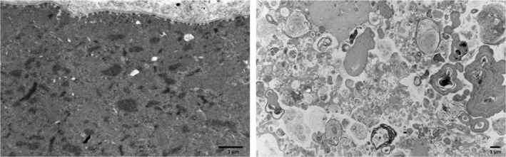
Background: myotilinopathy is a very rare inherited muscle disease that belongs to the group of myofibrillar myopathies. These diseases share a common alteration of the sarcomere organization at the level of the Z disk resulting in pathological protein aggregation, autophagic abnormalities, and ultimately muscle degeneration. Most reported cases are due to dominant missense mutations in the MYOT gene, two of which are largely recurrent.

Methods: We describe the clinical, radiological, pathological, and molecular analysis including long-read sequencing of a family affected by late-onset dominant proximodistal myopathy and muscle hypertrophy.

Results: We identified a duplication of the entire MYOT gene as the molecular cause of late-onset-myotilinopapthy with typical clinical and pathological features.

Conclusions: This study expands the molecular spectrum of myotilinopathy and highlights the use of long-read sequencing in the diagnosis of genetic neurological diseases caused by duplications and genomic structural variants. Myotilinopathy as well as other myofibrillar and distal myopathies should be considered in the differential diagnosis of patients affected by distal muscle weakness, even when presenting at an old age.

Keywords: distal myopathy; duplication; late‐onset myopathy; long‐read sequencing; muscle hypertrophy; myofibrillar myopathy; myotilin.


Conflict of interest statement

The authors declare no conflict of interest.

Figures

FIGURE 1
FIGURE 1
Molecular characterization and muscle imaging phenotype. (a) Pedigree of the family. Clinically affected patients are shown in black. Only the proband and his brother and sister could be tested for MYOT gene analysis. (b) Muscle MRI. In the proband (patient 1), severe fatty replacement is present in small glutei in the pelvis. In the thigh, fatty replacement shows a patchy asymmetrical distribution in posterior more than in anterior compartments, with severe involvement of adductor muscles, more patchy and asymmetrical involvement in biceps femoris, semimembranosus and semitendinosus. In the legs, soleus, medial gastrocnemius and peroneal muscles are involved. In the brother (patient 2), fatty replacement is present in small glutei bilaterally and middle and large gluteus on the left side. In the thigh patchy involvement of the left vastus lateralis and adductor muscles is present, while in the legs both soleus and to a lesser extent gastrocnemius are affected. (c) IGV screenshot of the proband's long‐read sequencing results of the duplicated region, including the entirety of MYOT as well as part of PKD2L2. The phased (haplotyped) alleles are shown in light blue and pink. The reads supporting the duplication (4 out of 20 and 4 out of 17 reads spanning the upstream and downstream breakpoints of the duplication, respectively) are shown in linked mode at the top of the alignment view. These supporting reads are characterized by their stretches of “rainbow” bases (arrows), which indicate that the reads, for those parts, map to another location in the genome than expected. By investigation of the sequences represented by these reads, the duplication was concluded to be in tandem. (d) Western blot analysis of MYOT protein expression of total muscle lysate of patient 1 compared to a pool of healthy controls. Myosin stained by Coomassie is the loading control. The patient and control pool were run on the same gel, the image was cropped for greater clarity.
FIGURE 2
FIGURE 2
Myopathological findings. (a) Light microscopy. Hematoxylin and eosin (H&E) staining shows increased fiber size variability with many hypertrophic fibers and some atrophic fibers, sometimes with an angular appearance. Some central nuclei are present and connective tissue content is increased. Cytosolic vacuoles, mostly non‐rimmed are indicated by black arrows. Basophilic material is evident in several fibers (white arrows). Similar findings are evident with Gomori trichrome staining (GT). NADH‐tetrazolium histoenzymatic reaction (NADH) shows abnormal myofibrillar network organization in both cases, with areas of variable size lacking reaction with core‐like lesions, and focal areas corresponding to the amorphous material indicated above showing intense reaction. Staining with anti‐myotilin antibody shows intense reaction of the protein deposits above described (MYOT). Strong reaction with anti‐p62 antibody in some muscle fibers. (b) Quantification of atrophic and hypertrophic muscle fibers. Atrophy factor is 274 for case 1 (n = 164 fibers analyzed) and 172 for case 2 (n = 533 fibers analyzed); hypertrophy factor was abnormally increased to 1884 for case 1 and 536 for case 2 [7].
FIGURE 3
FIGURE 3
Electron microscopy. Destruction of myofibrillar organization with accumulation of material derived from Z‐disk (left). Severe autophagic abnormalities and accumulation of degraded cytosolic material (right).

References

    1. Nakano S, Engel A, Waclawik J, Emslie‐Smith A, Busis N. Myofibrillar myopathy with abnormal foci of desmin positivity. I. Light and electron microscopy analysis of 10 cases. J Neuropathol Exp Neurol. 1996;55:549‐562. - PubMed
    1. Béhin A, Salort‐Campana E, Wahbi K, et al. Myofibrillar myopathies: state of the art, present and future challenges. Rev Neurol (Paris). 2015;171(10):715‐729. - PubMed
    1. Selcen D. Myofibrillar myopathies. Neuromuscul Disord. 2011;21(3):161‐171. - PMC - PubMed
    1. Olivé M, Goldfarb LG, Shatunov A, Fischer D, Ferrer I. Myotilinopathy: refining the clinical and myopathological phenotype. Brain. 2005;128(10):2315‐2326. - PubMed
    1. Chakravorty S, Nallamilli BRR, Khadilkar SV, et al. Clinical and genomic evaluation of 207 genetic myopathies in the Indian subcontinent. Front Neurol. 2020;11:1‐47. doi:10.3389/fneur.2020.559327 - DOI - PMC - PubMed

Publication types

LinkOut - more resources