Review
. 2025 Jan;240(1):e31500.
doi: 10.1002/jcp.31500.

Physiological Insights Into the Role of Pericytes in Spinal Cord Injury

Affiliations
Review

Physiological Insights Into the Role of Pericytes in Spinal Cord Injury

Haiming Jin et al. J Cell Physiol. 2025 Jan.

Abstract

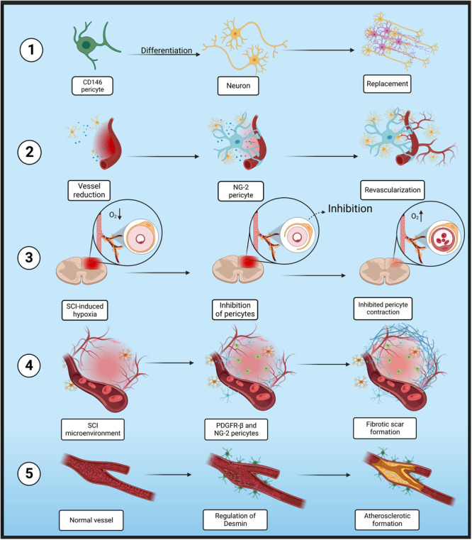
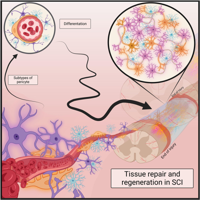
Vascular regeneration plays a vital role in tissue repair yet is drastically impaired in those with a spinal cord injury (SCI). Pericytes are of great significance as they are entwined with vessel-specific endothelial cells and actively contribute to maintaining the spinal cord's vascular network. Within the neurovascular unit (NVU), subtypes of pericytes characterized by various markers such as PDGFR-β, Desmin, CD146, and NG-2 are involved in vascular regeneration in SCI repair. Various pericyte signaling, pericyte-derived exosomes, and endothelial-pericyte interplay were revealed to participate in SCI repair or fibrotic scars. Through further understanding pericyte biology, it is aimed to accurately generate subtypes of pericytes and develop their therapeutic potential. This review focuses on recent advanced research and development of pericytes as a potential treatment for SCI.

Keywords: pericyte; physiology; spinal cord injury.


Conflict of interest statement

The authors declare no conflicts of interest.

Figures

Figure 1
Figure 1
The potential role of pericytes in SCI. (1) CD146 pericytes have demonstrated the ability to differentiate into neurocytes and replace damaged neurons. (2) During, SCI‐induced disregulation of angiogenesis, NG‐2 pericytes may promote revascularization, tissue healing, and vascular stability. (3) Inhibiting pericyte contraction has been found to effectively reduce localized tissue hypoxia subsequent to SCI. (4) In SCI microenvironment, PDGFR‐β pericytes may serve as a reservoir for scar‐forming cells, and together with NG‐2 pericytes, may contribute to scar formation. (5) Desmin‐type pericytes are known to be involved in the atherosclerotic formation through regulation of stem and endothelial cells.
Figure 2
Figure 2
Schematic representation of the localization, morphology, and coverage of various subtypes of pericytes and their potential in tissue regeneration in SCI.

Similar articles

References

    1. Alarcon‐Martinez, L. , Yilmaz‐Ozcan S., Yemisci M., et al. 2018. “Capillary Pericytes Express Alpha‐Smooth Muscle Actin, Which Requires Prevention of Filamentous‐Actin Depolymerization for Detection.” eLife 7: e34861. 10.7554/eLife.34861. - DOI - PMC - PubMed
    1. Bae, S. G. , Yin G. N., Ock J., Suh J. K., Ryu J. K., and Park J.. 2024. “Single‐Cell Transcriptome Analysis of Cavernous Tissues Reveals the Key Roles of Pericytes in Diabetic Erectile Dysfunction.” eLife 12: RP88942. - PMC - PubMed
    1. Baek, S. H. , Maiorino E., Kim H., Glass K., Raby B. A., and Yuan K.. 2022. “Single Cell Transcriptomic Analysis Reveals Organ Specific Pericyte Markers and Identities.” Frontiers in Cardiovascular Medicine 9: 876591. 10.3389/fcvm.2022.876591. - DOI - PMC - PubMed
    1. Bell, R. D. , Winkler E. A., Sagare A. P., et al. 2010. “Pericytes Control Key Neurovascular Functions and Neuronal Phenotype in the Adult Brain and During Brain Aging.” Neuron 68, no. 3: 409–427. - PMC - PubMed
    1. Buizza, C. , Enström A., Carlsson R., and Paul G.. 2024. “The Transcriptional Landscape of Pericytes in Acute Ischemic Stroke.” Translational Stroke Research 15, no. 4: 714–728. - PMC - PubMed

LinkOut - more resources