. 2024 Dec 10;12(1):ofae716.
doi: 10.1093/ofid/ofae716. eCollection 2025 Jan.

Diagnostic Impact of FISHseq as a New Pathologic Criterion for Endocarditis According to the Duke Criteria

Affiliations

Diagnostic Impact of FISHseq as a New Pathologic Criterion for Endocarditis According to the Duke Criteria

Alexander G M Hopf et al. Open Forum Infect Dis. .

Abstract

Background: For clinicians treating patients with infective endocarditis (IE), identifying the causative microorganisms poses a critical diagnostic challenge. Standard techniques including blood and heart valve cultures often yield inconclusive results. According to the recent 2023 Duke-ISCVID Criteria, molecular methods represent potent tools to enhance this aspect of IE diagnostics and guide subsequent therapeutic strategies.

Methods: We retrospectively analyzed data from 124 consecutive patients who underwent heart valve surgery due to suspected IE at München Klinik Bogenhausen. The standard diagnostic pathway, which included blood culture, valve culture, histopathological analysis, and polymerase chain reaction (PCR)/sequencing, was compared with the enhanced diagnostic pathway, which included fluorescence in situ hybridization + PCR/sequencing (FISHseq) instead of PCR/sequencing alone. The aim of this study was to assess the added value of combining standard diagnostics with molecular methods such as PCR/sequencing or FISHseq for the diagnosis of IE and the potential impact on therapy.

Results: Standard diagnostic methods and PCR/sequencing yielded inconclusive results in 57/124 cases (46.0%). FISHseq provided an added value for diagnostics in 79/124 cases (63.7%) and potentially would have impacted therapy in 95/124 (76.6%) of cases. By adding data through direct visualization and characterization of microorganisms, FISHseq reduced the number of inconclusive cases by 86.0%.

Conclusions: The comparison of 2 molecular diagnostic tools for IE from the same heart valve emphasizes the value of molecular methods including molecular imaging by FISH for IE diagnostics and supports the 2023 Duke-ISCVID Criteria.

Keywords: 2023 Duke-ISCVID Criteria; FISHseq; fluorescence in situ hybridization; infective endocarditis; molecular imaging.


Conflict of interest statement

Potential conflicts of interest. J.K. is CEO of MoKi Analytics GmbH. A.M. and J.K. are shareholders of MoKi Analytics GmbH. A.M. is the owner of the private practice Moter Diagnostics. All other authors report no potential conflicts.

Figures

Graphical Abstract
Graphical Abstract
https://tidbitapp.io/tidbits/diagnostic-impact-of-fishseq-as-a-new-pathologic-criterion-for-endocarditis-according-to-the-duke-criteria?utm_campaign=tidbitlinkshare&utm_source=IO
Figure 1.
Figure 1.
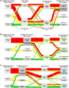
Reclassification of IE cases according to the modified Duke Criteria upon inclusion of postoperative valve culture and histopathology results (A), PCR/sequencing results (B), or FISHseq results (Enhanced Diagnostic Pathway) (C). Red indicates preoperative “definite IE,” yellow indicates “possible IE,” and green indicates “rejected IE.” A, Reclassification of IE cases according to the modified Duke Criteria after inclusion of postoperative VC and histopathology results. B, Reclassification of IE cases after inclusion of PCR/sequencing results (Standard Diagnostic Pathway). C, Reclassification of IE cases after inclusion of FISHseq results (Enhanced Diagnostic Pathway). Abbreviations: FISHseq, fluorescence in situ hybridization + polymerase chain reaction/sequencing; IE, infective endocarditis; MO, microorganism; PCR, polymerase chain reaction; VC, valve culture.
Figure 2.
Figure 2.
FISHseq workflow. After resection of heart valve tissue and fixation in FISHopt solution, the sample is embedded in cold polymerized resin. Subsequently, FISH is performed as outlined in Supplementary Data  C to enable an initial FISH report within 24 hours. Furthermore, DNA extraction from consecutive sample sections, as used for FISH, is used for PCR/sequencing. A final FISHseq report is usually available within 36–48 hours after surgery. Abbreviations: FISH, fluorescence in situ hybridization; FISHseq, fluorescence in situ hybridization + polymerase chain reaction/sequencing; PCR, polymerase chain reaction.
Figure 3.
Figure 3.
Standard and enhanced diagnostic pathway. Description of the standard and enhanced diagnostic pathway. The key difference of both pathways is that FISHseq is used instead of PCR/sequencing within the Enhanced Diagnostic Pathway. Overall, 124 patients underwent both pathways, but some patient samples did not undergo histopathology or valve culture. Abbreviations: FISHseq, fluorescence in situ hybridization + polymerase chain reaction/sequencing; PCR, polymerase chain reaction.
Figure 4.
Figure 4.
Discrepancy analysis of microbial genus/species identification from heart valve tissue samples using valve culture, PCR/sequencing, and FISHseq. Overall, in 77/124 patients a microbial genus/species was identified from heart valve tissue using valve culture, PCR/sequencing, or FISHseq. In 24/122 cases in which valve culture was performed, VC, PCR/sequencing, and FISHseq identified the same pathogen. In 36/122 cases in which VC was performed, VC failed to identify a pathogen, whereas PCR/sequencing identified a pathogen in 36/36 of these cases and FISHseq identified a pathogen in 32/36 of these cases. In 5/122 cases in which VC was performed, VC identified a different pathogen compared with PCR/sequencing and FISHseq. In 6/122 cases in which VC was performed and identified a pathogen with PCR/sequencing remaining negative, FISHseq identified a pathogen in 5/6 cases. In 6/122 cases in which VC was performed and both VC and PCR remained negative, FISHseq identified a new pathogen. In 2 cases, no VC was performed. In 45/122 cases where VC was performed, neither VC, PCR/sequencing, nor FISHseq identified a pathogen. Abbreviations: FISHseq, fluorescence in situ hybridization + polymerase chain reaction/sequencing; PCR, polymerase chain reaction; VC, valve culture.
Figure 5.
Figure 5.
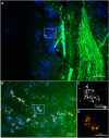
FISHseq detecting active infective endocarditis. FISH of a heart valve section from a 65-year-old male patient with an active IE caused by Gemella morbillorum. Blood cultures, PCR/sequencing, and FISHseq identified G. morbillorum, whereas the valve culture remained negative. FISHseq visualized active bacteria in biofilm formation. The heart valve section was stained using DAPI for visualization of nucleic acids in host cell nuclei and bacteria. A, Overview of the autofluorescent tissue background in green (enhanced for better orientation) with host cell nuclei and bacterial biofilms stained by DAPI in blue. The inset marks a region where bacterial biofilms are visible. B, At higher magnification, microorganisms stained with DAPI in blue and partly hybridized with the panbacterial probe EUB338Cy3 probe in orange are shown for visualization of active bacteria (based on ribosome content). The hybridization mix also contained NON338, the antisense probe of EUB388 as a nonsense control probe (not shown). The inset (C) shows at even higher magnification single bacteria that are partly FISH-positive and partly only DAPI-positive. C & D, The same field of view as with single separate microscopic channels for DAPI in black and white, and EUB338 in orange. Abbreviations: FISH, fluorescence in situ hybridization; FISHseq, fluorescence in situ hybridization + polymerase chain reaction/sequencing; PCR, polymerase chain reaction.
Figure 6.
Figure 6.
FISHseq showing treated/inactive infective endocarditis. FISH of a native heart valve section from a 64-year-old female patient, classified as “past IE,” where FISHseq visualized FISH-negative biofilms and identified E. faecalis in sequencing (confirmed by PCR/sequencing alone). Blood cultures had not been taken, and valve cultures remained negative. The section was hybridized with the panbacterial probe EUB338Cy3 for visualization of active bacteria (based on ribosome content), the nonsense control probe NON338Cy5, and stained with DAPI for visualization of nucleic acids in host cell nuclei and microorganisms. A, Overview shows the autofluorescent tissue background in green with DAPI-stained bacterial biofilms in blue. The inset marks the region magnified in (B), where DAPI only–positive bacteria are visible (overview of the channels: FITC—tissue background in green, Cy3—EUB338 probe in orange, Cy5—NON338 probe in magenta). The inset marks the region shown at higher magnification in (C–E). C–E, The same microscopic field of view with a merged image of all channels (C), the Cy3—EUB338 channel separately without any signal (D), and the Cy5—NON338 channel separately also without any signal (E). Abbreviations: FISH, fluorescence in situ hybridization; FISHseq, fluorescence in situ hybridization + polymerase chain reaction/sequencing; IE, infective endocarditis.
Figure 7.
Figure 7.
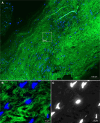
FISHseq showing no signs of infective endocarditis. FISH of a heart valve section from a 55-year-old male patient, initially suspected of IE. Neither the Standard nor Enhanced Diagnostics Pathway resulted in identification of a causative microorganism, with FISHseq visually not detecting any bacteria in the heart valve sample investigated. Hence, this patient was classified as “no IE.” The section was hybridized with the panbacterial probe EUB338Cy3 for visualization of active bacteria (based on ribosome content), the nonsense control probe NON338Cy5, and DAPI for visualization of nucleic acids in host cell nuclei and bacteria. A, Overview shows the autofluorescent heart valve tissue in green with host cell nuclei only in blue; no bacteria are visible at higher magnification (B and C). B, The merged image of the DAPI channel (blue) and the FITC channel in green. C, Greyscale image of the separate DAPI channel. No bacteria are visible. Abbreviations: FISH, fluorescence in situ hybridization; FISHseq, fluorescence in situ hybridization + polymerase chain reaction/sequencing; IE, infective endocarditis.
Figure 8.
Figure 8.
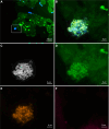
FISHseq proving infection with Staphylococcus epidermidis. FISH of a heart valve section from a 61-year-old male patient that was classified as possible IE using the modified Duke Criteria preoperatively but classified as inconclusive by the Standard Diagnostic Pathway because the histological examination of his heart valve showed signs of active IE, whereas all other methods were negative. In this case, FISHseq showed S. epidermidis in biofilms. A, The overview of the heart valve tissue shows the tissue background in green and bacterial biofilms at different locations within the tissue and at the tissue edges. The inset marked in (A) shows a region magnified in (B–F). The FISH was performed with the pan-bacterial probe EUB338FITC in green, the staphylococci genus–specific probe STAPHYCy3 in orange, the nonsense probe NON338Cy5 as negative binding control in magenta, and the nucleic acid stain DAPI. B, A merged image of all channels. C–F, The same microscopic field of view with separate channels for the nucleic acid stain DAPI in black and white, the EUB338 probe and the tissue background in green, the STAPHY probe in orange, and the negative control probe in magenta, respectively. Visible are active staphylococci visualized by the pan-bacterial FISH probe EUB338 and the staphylococci genus–specific FISH probe STAPHY, whereas no signal is detectable with the nonsense probe NON338. Abbreviations: FISH, fluorescence in situ hybridization; FISHseq, fluorescence in situ hybridization + polymerase chain reaction/sequencing; IE, infective endocarditis.
Figure 9.
Figure 9.
Potential impact of FISHseq results on antibiotic therapy. The figure shows the potential impact of FISHseq results on the antibiotic therapy of the IE patients in this study. The crimson color indicates conclusively positive patients after FISHseq evaluation; blue indicates inconclusive patients, and green indicates conclusively negative patients. The possible impact on antibiotic therapy is based on current guidelines and selected publications and refers to a potential therapy escalation because of microbial biofilms in the heart valve, a targeted antibiotic therapy upon causative pathogen identification, or therapy de-escalation because of inactive bacteria/absence of bacteria. Abbreviations: FISHseq, fluorescence in situ hybridization + polymerase chain reaction/sequencing; IE, infective endocarditis.

References

    1. Liesman RM, Pritt BS, Maleszewski JJ, Patel R. Laboratory diagnosis of infective endocarditis. J Clin Microbiol 2017; 55:2599–608. - PMC - PubMed
    1. Cahill TJ, Baddour LM, Habib G, et al. Challenges in infective endocarditis. J Am Coll Cardiol 2017; 69:325–44. - PubMed
    1. Coffey S, Roberts-Thomson R, Brown A, et al. Global epidemiology of valvular heart disease. Nat Rev Cardiol 2021; 18:853–64 - PubMed
    1. Li JS, Sexton DJ, Mick N, et al. Proposed modifications to the Duke Criteria for the diagnosis of infective endocarditis. Clin Infect Dis 2000; 30:633–8. - PubMed
    1. Fowler VG, Durack DT, Selton-Suty C, et al. The 2023 Duke-ISCVID Criteria for infective endocarditis: updating the modified Duke Criteria. Clin Infect Dis 2023; 77:518–26. - PMC - PubMed