. 2024 Sep 19;27(11):110989.
doi: 10.1016/j.isci.2024.110989. eCollection 2024 Nov 15.

Disrupting the transmembrane domain interface between PMP22 and MPZ causes peripheral neuropathy

Affiliations

Disrupting the transmembrane domain interface between PMP22 and MPZ causes peripheral neuropathy

Natalya Pashkova et al. iScience. .

Abstract

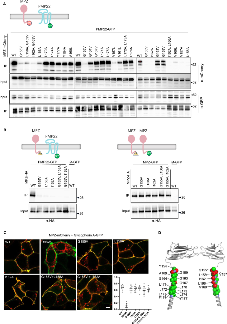
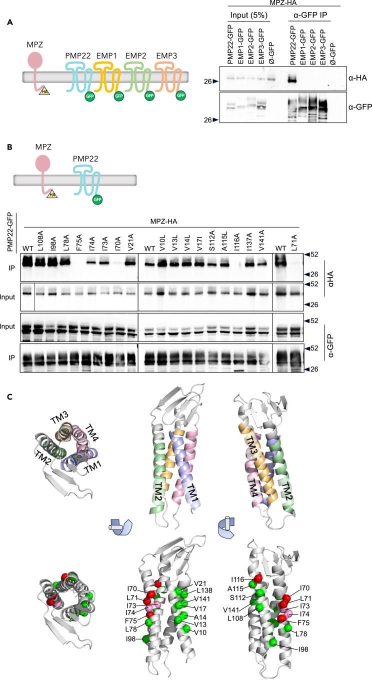
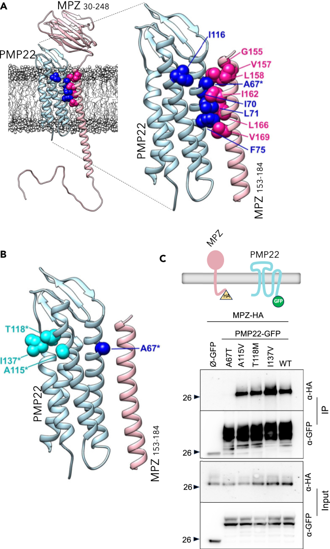
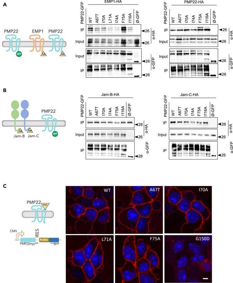
Peripheral Myelin Protein 22 (PMP22) and MPZ are abundant myelin membrane proteins in Schwann cells. The MPZ adhesion protein holds myelin wraps together across the intraperiod line. PMP22 is a tetraspan protein belonging to the Claudin superfamily. Loss of either MPZ or PMP22 causes severe demyelinating Charcot-Marie-Tooth (CMT) peripheral neuropathy, and duplication of PMP22 causes the most common form of CMT, CMT1A. Yet, the molecular functions provided by PMP22 and how its alteration causes CMT are unknown. Here, we find MPZ and PMP22 form a specific complex through interfaces within their transmembrane domains. We also find that the PMP22 A67T patient variant that causes a loss-of-function (hereditary neuropathy with pressure palsies) phenotype maps to this interface, and blocks MPZ association without affecting localization to the plasma membrane or interactions with other proteins. These data define the molecular basis for the MPZ ∼ PMP22 interaction and indicate this complex fulfills an important function in myelinating cells.

Keywords: Biochemistry; Cell biology; Molecular biology; Neuroscience.


Conflict of interest statement

The authors declare there are no competing interests.

Figures

None
Graphical abstract
Figure 1
Figure 1
MPZ and PMP22 form a complex in HEK293 and RT4 Schwannoma cells (A) Left panel shows scheme for co-immunoprecipitation in which MPZ carrying a C-terminal HA tag and PMP22 carrying a C-terminal GFP tag are co-expressed and then immunoprecipitated with Sepharose linked to α-GFP nanobodies. Right panel shows results of co-immunoprecipitation. Lysates from HEK293 cells transiently co-transfected with MPZ-HA and PMP22-GFP were prepared with 0.5% NP-40 and 0.5% Triton X-100 or with 1% DDM and 0.2% CHS detergents. Bead-bound precipitates were washed in PBS with their respective detergents and eluted with SDS sample buffer. A proportion of the input (5%) was analyzed along with precipitated complexes by SDS-PAGE and immunoblotting with mouse α-HA (to detect associated MPZ-HA) and mouse α-GFP (to show efficiency of PMP22-GFP immunoprecipitation). (B) The co-immunoprecipitation scheme in A was used for HEK293 cells in which PMP22-GFP and MPZ-HA or MPZ-GFP and MPZ-HA were co-expressed together before (pre) lysis or were expressed in separate populations and mixed after lysis (post). Immunoprecipitation samples together with a 10% equivalent of the starting input was immunoblotted with anti-HA to reveal associated MPZ-HA. Note that while MPZ-HA associates with MPZ-GFP, the level of this association is far less than that with PMP22-GFP. Moreover, both associations required that proteins had to be expressed in the same cells prior to detergent lysis to detect the presence of MPZ ∼ MPZ or MPZ ∼ PMP22 complexes. (C) Co-immunoprecipitation of PMP22-GFP and MPZ-HA from DDM/CHS detergent lysates of transiently transfected RT4 Schwannoma cells. Whereas MPZ-HA was co-precipitated with PMP22-GFP, it was not co-precipitated with the PMP22-related protein EMP3-GFP. (D) Top panel shows scheme for co-immunoprecipitation of MPZ-GFP with PMP22 containing an exofacial myc epitope tag inserted into the second extracellular loop. Bottom panel shows PMP22-mycexo is recovered when co-expressed with MPZ-GFP and immunoprecipitated with α-GFP nanobody beads. (E) Quantitation of FRET efficiency between PMP22-GFP and MPZ-mCherry and between MPZ-GFP and MPZ-mCherry. FRET was measured as an increase in donor fluorescence after acceptor photobleaching in transiently transfected HEK293 cells. The “positive” control was MPZ tandemly fused to both msGFP2 and mCherry. The “negative” control was Glycophorin A-GFP and MPZ-mCherry. Regions of interest were analyzed and quantified for at least 25 cells from 2 different experiments. FRET efficiencies were calculated with the Leica LAS X Microlab module. All ROI data are represented singly and aggregated as mean FRET efficiency ±S.D (from left to right 0.237 ± 0.052, 0.014 ± 0.020, 0.079 ± 0.042, 0.053 ± 0.029). One-way ANOVA with Turkey’s multiple comparison test revealed the negative control was significantly different from each of the other samples. On the right is a representative FRET image of PMP22-GFP + MPZ-mCherry. The yellow boxes on the images show regions of interest used for bleaching and FRET measurements. Bar = 5μm.
Figure 2
Figure 2
Specificity of MPZ association with PMP22 (A) Several members of the MPZ/β-subunit family were assessed for their ability to associate with PMP22-GFP using the co-immunoprecipitation scheme outlined in Figure 1. DDM/CHS detergent lysates from HEK293 cells expressing the indicated HA-tagged MPZ/β-subunit proteins with either PMP22-GFP or EMP3-GFP were immunoprecipitated with α-GFP nanobody beads. Immunoprecipitation of PMP22-GFP only efficiently recovered MPZ-HA and a very low level of MPZL1. In contrast, MPZL2, SCN3B, and SCN2B formed strong complexes with EMP3-GFP. A 5% equivalent of the input lysate was included in each immunoblot. (B) The indicated HA-epitope tagged chimeric proteins made of the extracellular Ig-like domain, TMD, and cytosolic domains of MPZL1, SCN2B, and MPZ were assessed for association with PMP22-GFP, EMP3-GFP, or GFP alone (Ø-GFP) as described in (A). Chimeras containing the TMD of MPZ were all co-precipitated with PMP22-GFP whereas substitution of the extracellular domain or cytosolic domain of MPZ with that of SCN2B or MPZL1 did not block PMP22 association. In contrast, the ability of SCN2B to associate with EMP3-GFP is conferred by its extracellular Ig-like containing domain.
Figure 3
Figure 3
Mapping the PMP22-interacting interface within the MPZ transmembrane segment (A) Top panel shows experimental scheme in which PMP22-GFP is co-expressed with mutant versions of MPZ-mCherry and immunoprecipitated with α-GFP nanobody beads. Lower panel shows immunoprecipitation results on the indicated MPZ-mCherry mutants. (B) Left. Results from A were verified using MPZ mutants in the context of an MPZ C-terminally HA-epitope-tagged protein co-expressed with PMP22-GFP or GFP alone (∅-GFP) by coimmunoprecipitation with α-GFP nanobody beads and immunoblotting with α-HA antibodies. Right. MPZ-HA Wild-type (WT) and the indicated mutants were assessed for their ability to co-immunoprecipitate with MPZ-GFP or GFP alone (Ø-GFP) using α-GFP nanobody beads. (C) Cell surface localization of WT MPZ-mCherry and the indicated mutants was assessed by confocal microscopy. Micrographs show WT MPZ-mCherry and the the indicated mutants at the plasma membrane marked by glycophorin A tagged with GFP. The MPZ R98W mutant, which is known to be retained in the ER, was included for comparison. Multiple fields encompassing >100 cells were assessed for overlap of MPZ-mCherry with the plasma membrane marker glycophorin A using Manders overlap coefficient and plotted for each field in graph at right along with mean ± SD. One way ANOVA revealed no differences in cell surface localization (p > 0.1) among mutants and the WT MPZ-mCherry with the exception of the R98W mutant that had a significantly lower cell surface localization (p < 0.0001) to every other MPZ-mCherry protein analyzed. Bar = 5μm. (D) Residues in the MPZ transmembrane segment that when mutated had no effect (green) and dramatic reduction (red) in the ability to associate with PMP22 were mapped onto a model (AlphaFold: AF-P25189-F1) of a portion of MPZ containing the extracellular Ig-like domain and the α-helical region encompassing the transmembrane segment. Note that the red residues align along one face of the α-helix forming a cluster indicating the likely interface mediating PMP22 association.
Figure 4
Figure 4
Specificity of PMP22 interaction with MPZ (A) PMP22-GFP or the PMP22-related proteins EMP1-GFP, EMP2-GFP, EMP3-GFP, or GFP alone (Ø-GFP) were co-expressed with MPZ-HA in HEK293 cells and immunoprecipitated with α-GFP nanobodies. Immunoprecipitated eluted proteins were immunoblotted with α-HA to detect associated MPZ-HA and α-GFP to monitor recovery of PMP22-GFP. A 5% equivalent of the input lysate was included in the immunoblots. (B) The indicated amino acid substitutions were incorporated into PMP22-GFP and assessed for association with co-expressed MPZ-HA. (C) Model of PMP22 (AlphaFold: AF-Q01453-F1) showing orientation of the four transmembrane helices (TM1-4) of PMP22 and the predicted position of the amino acids critical for MPZ binding. Residues in PMP22 that when mutated had no effect (green), a partial reduction (pink) and dramatic reduction (red) in the ability to associate with MPZ are shown.
Figure 5
Figure 5
The A67T patient variant of PMP22 causing an HNPP phenotype maps to the presumed interface between PMP22 and MPZ (A) Model of the complex between PMP22 and MPZ in which association is mediated by residues identified in amino acid substitution experiments. (Upper panel) The multi-component model composed of the full-length human PMP22 (light blue—AlphaFold: AF-Q01453-F1) and MPZ (pink—AlphaFold: AF-P25189-F1) is oriented in a lipid bilayer of 60% POPC: 40% cholesterol (white). For a few unstructured residues between MPZ domains, torsion adjustments were made for improved visualization of the extracellular domain and the flexible portion of the cytosolic tail. The residues that destroy or diminish the association of PMP22 and MPZ when mutated are indicated. The position of the HNPP-causing patient variant, A67T, which lies within the predicted interface mediating MPZ ∼ PMP22 binding is indicated (∗). (B) Model of the PMP22∼MPZ interface shown in (A) with the positions of select patient variants including the A67T indicated on the different transmembrane helices. (C) Effect of PMP22 patient variants on the association with MPZ. MPZ-HA was co-expressed with GFP alone (Ø-GFP), WT PMP22-GFP or the indicated PMP22 patient variants and assessed for complex assembly by coimmunoprecipitation with α-GFP nanobody beads and immunoblotting for MPZ-HA with α-HA antibodies. Comparable expression levels of the different PMP22 patient variants as well as the efficiency were assessed by immunoblotting input lysates and bead immunoprecipitates with α-GFP antibodies.
Figure 6
Figure 6
Specificity of PMP22 mutants lacking the ability to bind MPZ (A) PMP22-GFP, both WT and the indicated mutants along with GFP alone (Ø-GFP), were assessed for their ability to bind EMP1-HA (left) or PMP22-HA (right). Comparable expression levels of the different PMP22 mutants as well as the efficiency was assessed by immunoblotting input lysates and bead immunoprecipitates with α-GFP antibodies. (B) PMP22-GFP, both WT and the indicated mutants, were assessed for the ability to bind Jam-B-HA (left) and Jam-C (right). (C) The cell surface localization of PMP22 WT and the indicated mutants was assessed in the context of the PMP22-mycexo protein containing an extracellular (exofacial) myc epitope. Expression of PMP22-mycexo was coupled to the expression of a nuclear-localized puromycin-blue fluorescent protein fusion by having it downstream of an internal ribosome entry site on the PMP22 mRNA. Stable transfected HEK293 cell lines expressing these mutants were fixed, left unpermeablized and labeled with α-myc monoclonal antibody and Alexa 568 secondary antibody prior to visualization by confocal microscopy (right). Bar = 5μm.
Figure 7
Figure 7
Immunogold labeling of MPZ and PMP22 in HEK cell freeze fracture replicas (A) Electron micrographs of the replicas from HEK293 cells expressing MPZ-mCherry alone (A) or (B–E) micrographs of HEK293 cells expressing both MPZ-mCherry and PMP22-GFP. Cells were allowed to form cell-cell contacts before fixation in 1% paraformaldehyde. ΜPZ-mCherry was labeled with 6 nm immunogold particles and PMP22-GFP was labeled with 12 nm immunogold particles. Where two cell membranes are contacting, the immunogold labeling for both proteins were observed exclusively on the protoplasmic face/leaflet (P-face) and always homogeneously spread throughout the plasma membrane rather than distributed in clusters. The labeling pattern was same in the single expression and labeling (A) and double expression and labeling (B). The topology of two contacting membrane is presentenced in (C). The labeled P-face shows continuity to its cytoplasm and an extraplasmic face/leaflet (E-face) of another cell is closely contacting on the P-face. The P-face exclusive labeling indicates the specificity of the labeling and consistent with the intercellular epitope locations. There were instances of intramembrane particles in a small cluster and strings observed on the E-face (D, E, arrows), but these were only rarely found. The small amount of immunogold particles near the string in (E) indicated the labeling of a hidden P-face below the E-face, called cryptic labeling, however the positions of the immunogold particles didn’t follow the string shape and could not be verified if they correspond to the localization of PMP22-GFP or MPZ-mCherry.

Update of

References

    1. Skre H. Genetic and clinical aspects of Charcot-Marie-Tooth's disease. Clin. Genet. 1974;6:98–118. - PubMed
    1. Fridman V., Bundy B., Reilly M.M., Pareyson D., Bacon C., Burns J., Day J., Feely S., Finkel R.S., Grider T., et al. CMT subtypes and disease burden in patients enrolled in the Inherited Neuropathies Consortium natural history study: a cross-sectional analysis. J. Neurol. Neurosurg. Psychiatry. 2015;86:873–878. doi: 10.1136/jnnp-2014-308826. - DOI - PMC - PubMed
    1. Murphy S.M., Laura M., Fawcett K., Pandraud A., Liu Y.T., Davidson G.L., Rossor A.M., Polke J.M., Castleman V., Manji H., et al. Charcot-Marie-Tooth disease: frequency of genetic subtypes and guidelines for genetic testing. J. Neurol. Neurosurg. Psychiatry. 2012;83:706–710. doi: 10.1136/jnnp-2012-302451. - DOI - PMC - PubMed
    1. Lemke G., Axel R. Isolation and sequence of a cDNA encoding the major structural protein of peripheral myelin. Cell. 1985;40:501–508. - PubMed
    1. Uyemura K., Asou H., Takeda Y. Structure and function of peripheral nerve myelin proteins. Prog. Brain Res. 1995;105:311–318. - PubMed

LinkOut - more resources