. 2024 Dec 20:15:1499471.
doi: 10.3389/fimmu.2024.1499471. eCollection 2024.

Targeting CEA in metastatic triple negative breast cancer with image-guided radiation followed by Fab-mediated chimeric antigen receptor (CAR) T-cell therapy

Affiliations

Targeting CEA in metastatic triple negative breast cancer with image-guided radiation followed by Fab-mediated chimeric antigen receptor (CAR) T-cell therapy

Eric Aniogo et al. Front Immunol. .

Abstract

Introduction: Although CAR-T cell therapy has limited efficacy against solid tumors, it has been hypothesized that prior treatment with Image-Guided Radiation Therapy (IGRT) would increase CAR-T cell tumor infiltration, leading to improved antigen specific expansion of CAR-T cells.

Methods: To test this hypothesis in a metastatic triple negative breast cancer (TNBC) model, we engineered two anti-CEA single-chain Fab (scFab) CAR-T cells with signaling domains from CD28zeta and 4-1BBzeta, and tested them in vitro and in vivo.

Results: The anti-CEA scFab CAR-T cells generated from three different human donors demonstrated robust in vitro expression, expansion, and lysis of only CEA-positive TNBC cells, with the CD28z-CAR-T cells showing the highest cytotoxicity. IFN-γ and granzyme B release assays revealed significantly higher IFN-γ production at a 4:1 effector-to-target (E:T) ratio in CD28z-CAR-T cells compared to 4-1BBz-CAR-T cells. Treatment of CEA-positive TNBC MDA-MB231 xenografts in the mammary fat pads of NSG mice, that produced spontaneous lung metastases over time, resulted in significant tumor growth reduction compared to either therapy alone (p<0.01). Immunohistochemical (IHC) analysis revealed that only combined IGRT and CAR-T therapy resulted in the elimination of lung metastases.

Discussion: These findings demonstrate that the combination of IGRT and anti-CEA scFab CAR-T therapy induces a strong antitumor response, effectively targeting both the primary tumor and distant metastatic lesions in the lungs, thus demonstrating that IGRT enhances CAR-T cell infiltration, persistence, and overall efficacy within both primary and metastatic lesions.

Keywords: carcinoembryonic antigen; cell immunotherapy; chimeric antigen receptor T cells; image-guided radiation therapy; triple-negative breast cancer.


Conflict of interest statement

The authors declare that the research was conducted in the absence of any commercial or financial relationships that could be construed as a potential conflict of interest. The author(s) declared that they were an editorial board member of Frontiers, at the time of submission. This had no impact on the peer review process and the final decision.

Figures

Figure 1
Figure 1
Schematics of humanized anti-CEA (M5A) scFab-CAR-T. (A) 4-1BB and (B) CD28 CAR constructs. Two constructs were tested, with cytoplasmic domain corresponding to second generation CAR-Ts. (C, D) The two scFab CAR-T constructs have distinct signaling domains; CD28-CD3zeta and 4-1BB-CDzeta configuration, both co-expressing hCD19t to allow identification of expressed CARs.
Figure 2
Figure 2
scFab-CAR-T production and expression on T cells. (A) Transduction efficiency was assessed by flow cytometry for CD19t expression of transduced T cells following enrichment with anti-CD19 beads. (B) Percentage expression of scFab-CAR-T cell production across T cells from three different donors. (C) Growth curves showing the expansion of scFab-CAR-T from three donors’ post-transduction.
Figure 3
Figure 3
In Vitro scFab-CAR T-cell cytotoxic assays; (A) Cytotoxicity against CEA-negative MDA-MB231 parental cells. (B) Cytotoxicity against CEA-positive MDA-MB231 cells. Both versions of scFab CAR-T cells exhibit antigen-specific lysis of target cells. (C, D) Analysis of IFN-γ and granzyme B production by mock and two versions of scFab CAR-T cells against CEA-positive MDA-MB231 cells. (E) Expression of the CD107a degranulation marker in both CAR-T cells when co-incubated with CEA-positive and negative MDA-MB231 target cells. (Statistical analysis was performed using two-way ANOVA and Sidak’s multiple comparison test *p < 0.05, **p < 0.01, ***p < 0.001, ****p < 0.0001, ns - not significant).
Figure 4
Figure 4
In Vivo therapeutic Efficacy of Anti-CEA scFab-CD28-CAR-T in combination with 10 Gy of IGRT. (A) Experimental design: orthotopic MDA-MB-231CEA-Luc positive tumors implanted in NSG mice were treated with 10 Gy IGRT on day 10, followed by an intravenous injection of 1 × 10⁶ anti-CEA scFab CAR-T cells on day 11. Tumor size was monitored with calipers until reaching 1500 mm³ (for controls and CAR T groups). (B) MDA-MB-231CEA-Luc tumor growth curves (n=4-5 mice per group). The combination therapy of 10 Gy IGRT and scFab-CAR-T cells was statistically significant (p<0.01). (C) Tumor weight measurements after euthanasia on day 48 showed a significant difference (p<0.0001) in tumor weight between the control and combined treatment groups. (D) Tumor analysis for T cell subpopulations (CD4/CD8) using flow cytometry and their corresponding expression of exhaustion markers in CD4 (E) and CD8 (F) subpopulations. (Statistical analysis was performed using two-way ANOVA and Sidak’s multiple comparison test *p < 0.05, **p < 0.01, ***p < 0.001, ****p < 0.0001).
Figure 5
Figure 5
Comparative immunohistochemistry analysis of infiltrating anti-CEA CAR-T cells in MDA-MB231CEA-Luciferase Tumors with or without IGRT. NSG mice with orthotopic MDA-MB-231CEA-Luciferase-positive tumors were analyzed as (A) Tumors treated with CAR-T cells only and (B) Tumors receiving combination treatment of 10Gy IGRT and anti-CEA CAR-T cells. Tumors were monitored until they reached terminal size or until day 48, and then analyzed using immunohistochemistry. Anti-CD3 staining indicates infiltrating anti-CEA CAR-T cells, that were significantly increased in the combination therapy group. (C) Quantification of infiltrating CD3-positive T cells in MDA-MB-231CEA-Luciferase tumors (2 regions per tumor, 5 tumors each group) showed a significant increase in the combination treatment group compared to the anti-CEA CAR-T cell-only group (p < 0.01). (Statistical analysis was performed using student t-test **p < 0.01).
Figure 6
Figure 6
Immunohistochemistry Staining of MDA-MB231CEA-Luc Tumor Metastasis to the Lungs. At termination, one lung lobe from each mouse was collected and stained to detect Luc+ tumor cells (A-D). (E) Quantification of Luc+ areas (average of 5 areas for each lobe) revealed a significant difference between control, single CAR-T treatment, and combination treatment groups (p < 0.001). Each group consisted of 4-5 mice. (Statistical analysis was performed using one-way ANOVA and Sidak’s multiple comparison test *p < 0.05, ***p < 0.001).
Figure 7
Figure 7
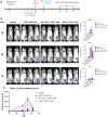
In vivo analysis of CAR-T cell tracking kinetics. (A) the treatment schedule schematic, (B) representative bioluminescence scans for CAR-T cell luciferase activity, and (C) the graphical quantification of luciferase in photons per second across the treatment groups. The untreated control group included 2 mice, while the other groups consisted of 3-5 mice each. (Statistical analysis was performed using two-way ANOVA and Sidak’s multiple comparison test **p < 0.01, ns - not significant).

References

    1. Alam MS, Rahaman MM, Sultana A, Wang G, Mollah MNH. Medicine: Statistics and network-based approaches to identify molecular mechanisms that drive the progression of breast cancer. Comput Biol Med. (2022) 145:105508. doi: 10.1016/j.compbiomed.2022.105508 - DOI - PubMed
    1. Yao H, He G, Yan S, Chen C, Song L, Rosol TJ, et al. . Triple-negative breast cancer: is there a treatment on the horizon? Oncotarget. (2017) 8:1913. doi: 10.18632/oncotarget.12284 - DOI - PMC - PubMed
    1. Bezerra P, Motti EF. 3-NAntC: A potent crotoxin B-derived peptide against the triple-negative MDA-MB-231 breast cancer cell line. Molecules. (2024) 29:1646. doi: 10.3390/molecules29071646 - DOI - PMC - PubMed
    1. Sanchez-Barcelo EJ, Mediavilla MD, Alonso-Gonzalez C, Reiter R. Melatonin uses in oncology: breast cancer prevention and reduction of the side effects of chemotherapy and radiation. Expert Opin Investig Drugs. (2012) 21:819–31. doi: 10.1517/13543784.2012.681045 - DOI - PubMed
    1. Adams S, Gatti-Mays ME, Kalinsky K, Korde LA, Sharon E, Amiri-Kordestani L, et al. . Current landscape of immunotherapy in breast cancer: a review. JAMA Oncology. (2019) 5:1205–14. doi: 10.1001/jamaoncol.2018.7147 - DOI - PMC - PubMed

MeSH terms

Substances

LinkOut - more resources