Cholangitis and Congestive Heart Failure Secondary to Biliary Hemorrhage in Hereditary Hemorrhagic Telangiectasia
- PMID: 39759662
- PMCID: PMC11700518
- DOI: 10.7759/cureus.75232
Cholangitis and Congestive Heart Failure Secondary to Biliary Hemorrhage in Hereditary Hemorrhagic Telangiectasia
Abstract
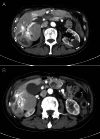
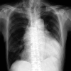
This case report discusses the case of a 74-year-old man who was diagnosed with hereditary hemorrhagic telangiectasia (HHT). The patient initially presented with right upper quadrant abdominal pain and was later diagnosed with cholangitis. Subsequently, heart failure was identified due to hepatic arteriovenous malformations. Although the exact duration of his disease remains unclear, the patient was initially asymptomatic and developed significant complications over time, possibly reflecting the progressive nature of vascular malformations associated with HHT. This case underscores the necessity of regular imaging follow-ups to assess the progression of vascular malformations in various organs, highlighting the clinical significance of the early detection and management of complications in HHT.
Keywords: abdominal pain; cholangitis; congestive heart failure; early detection; hepatic arteriovenous malformations; hereditary hemorrhagic telangiectasia.
Copyright © 2024, Yamamoto et al.
Conflict of interest statement
Human subjects: Consent for treatment and open access publication was obtained or waived by all participants in this study. Conflicts of interest: In compliance with the ICMJE uniform disclosure form, all authors declare the following: Payment/services info: All authors have declared that no financial support was received from any organization for the submitted work. Financial relationships: All authors have declared that they have no financial relationships at present or within the previous three years with any organizations that might have an interest in the submitted work. Other relationships: All authors have declared that there are no other relationships or activities that could appear to have influenced the submitted work.
Figures


References
-
- Age-related clinical profile of hereditary hemorrhagic telangiectasia in an epidemiologically recruited population. Plauchu H, de Chadarévian JP, Bideau A, Robert JM. Am J Med Genet. 1989;32:291–297. - PubMed
-
- Genetic epidemiology of hereditary hemorrhagic telangiectasia in a local community in the northern part of Japan. Dakeishi M, Shioya T, Wada Y, et al. Hum Mutat. 2002;19:140–148. - PubMed
-
- Hereditary haemorrhagic telangiectasia: a population-based study of prevalence and mortality in Danish patients. Kjeldsen AD, Vase P, Green A. J Intern Med. 1999;245:31–39. - PubMed
-
- Doppler ultrasonographic grading of hepatic vascular malformations in hereditary hemorrhagic telangiectasia -- results of extensive screening. Buscarini E, Danesino C, Olivieri C, et al. Ultraschall Med. 2004;25:348–355. - PubMed
-
- Hereditary hemorrhagic telangiectasia: multi-detector row helical CT assessment of hepatic involvement. Ianora AA, Memeo M, Sabba C, Cirulli A, Rotondo A, Angelelli G. Radiology. 2004;230:250–259. - PubMed
Publication types
LinkOut - more resources
Full Text Sources
