. 2024 Dec 10:46:97-117.
doi: 10.1016/j.bioactmat.2024.12.008. eCollection 2025 Apr.

A deformable SIS/HA composite hydrogel coaxial scaffold promotes alveolar bone regeneration after tooth extraction

Affiliations

A deformable SIS/HA composite hydrogel coaxial scaffold promotes alveolar bone regeneration after tooth extraction

Shiqing Ma et al. Bioact Mater. .

Abstract

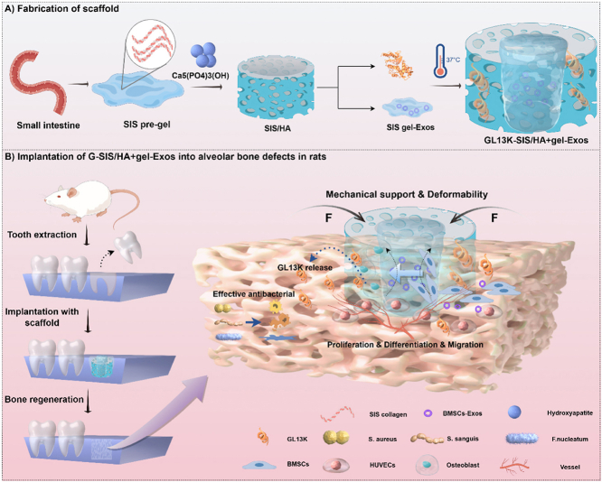
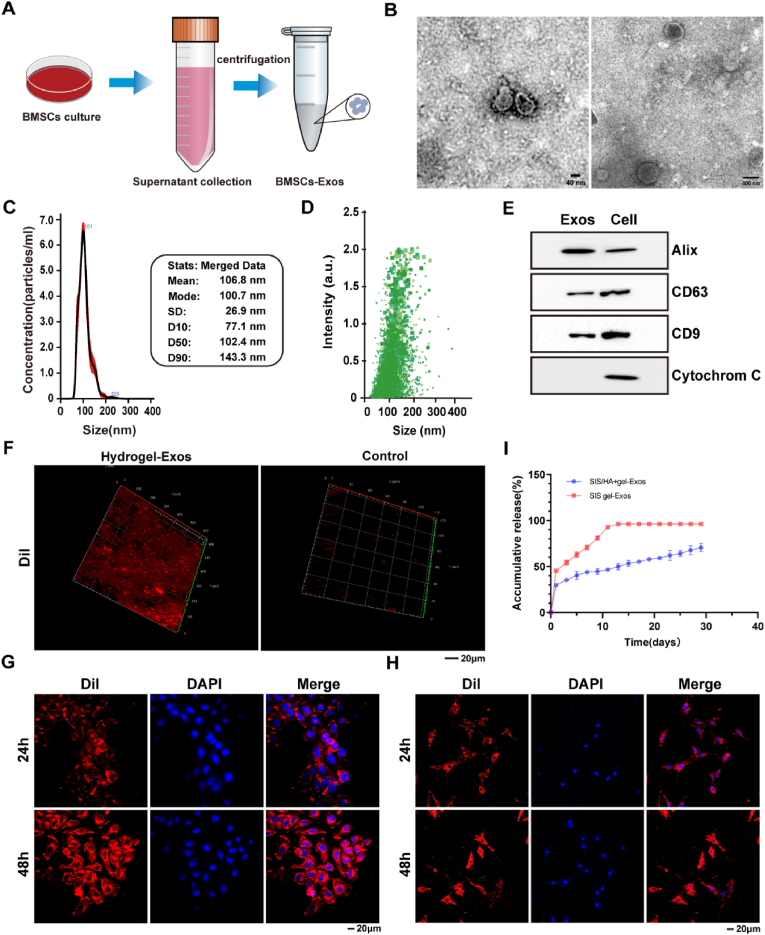
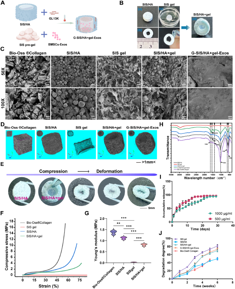
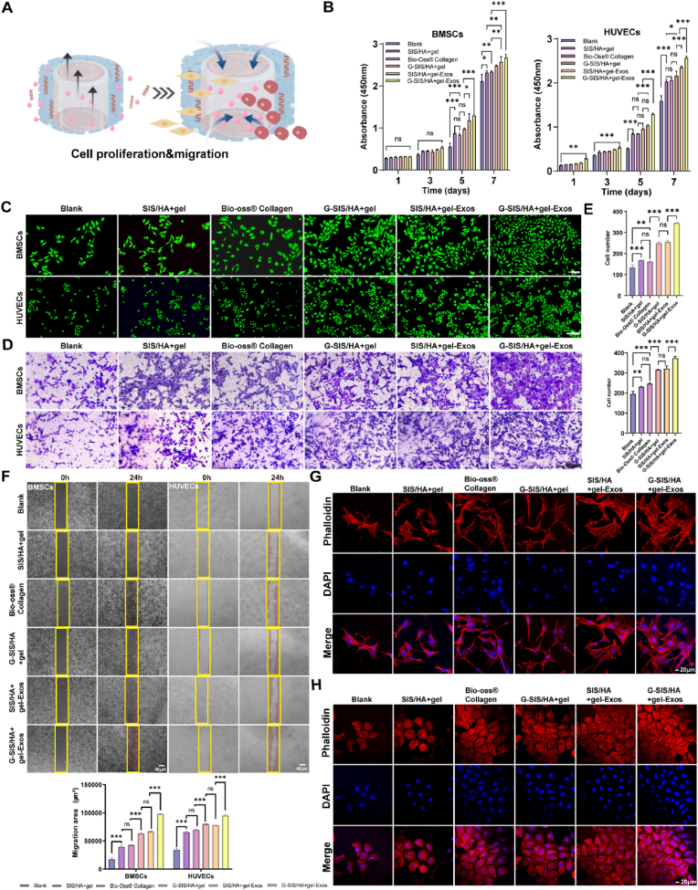
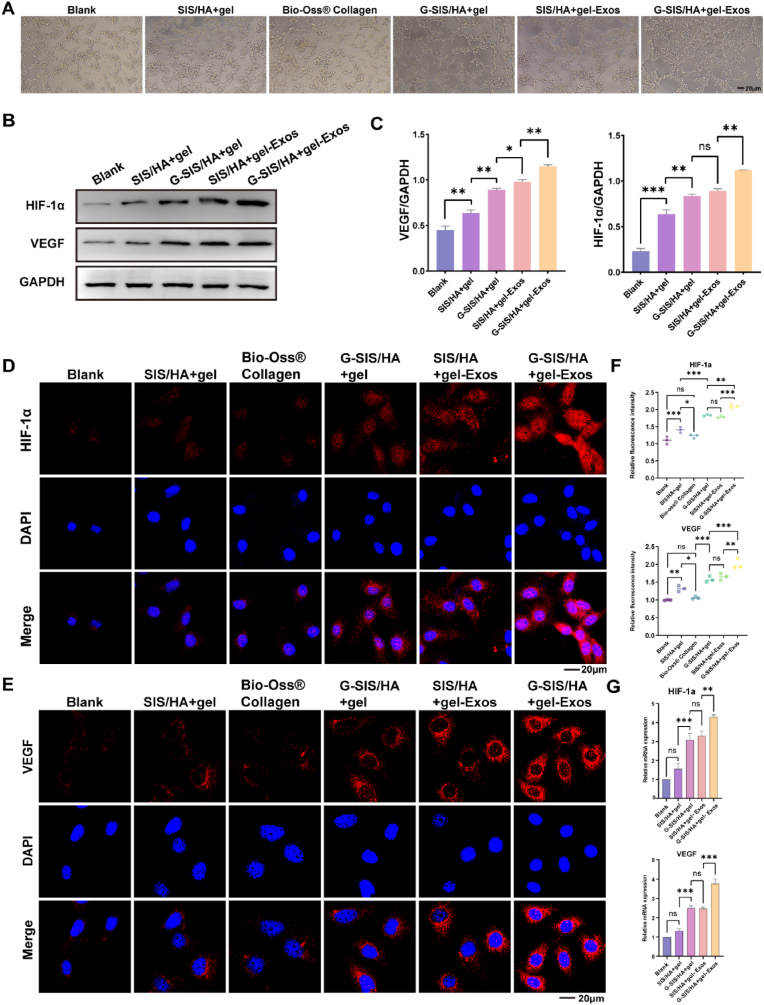
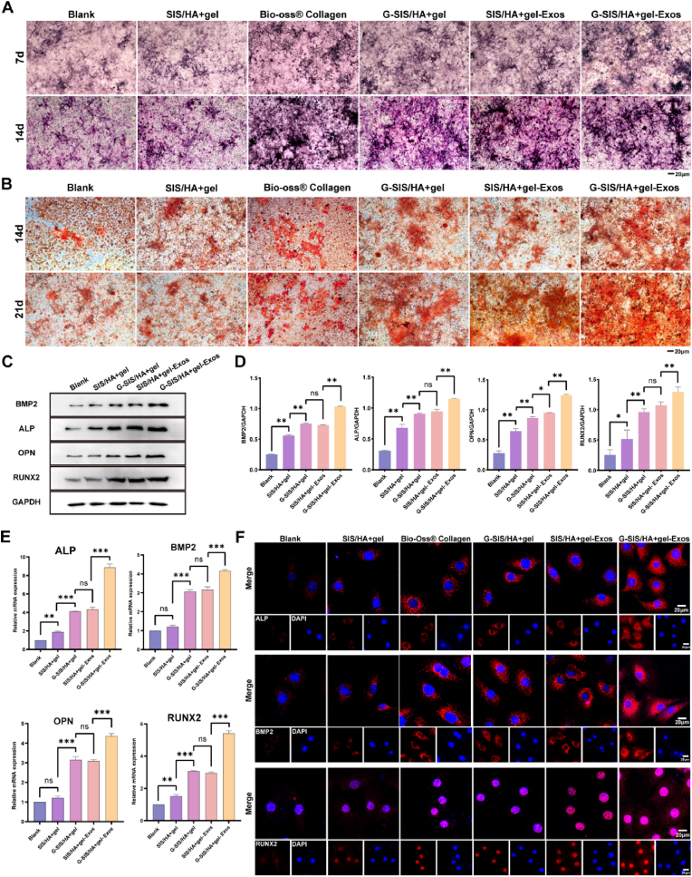
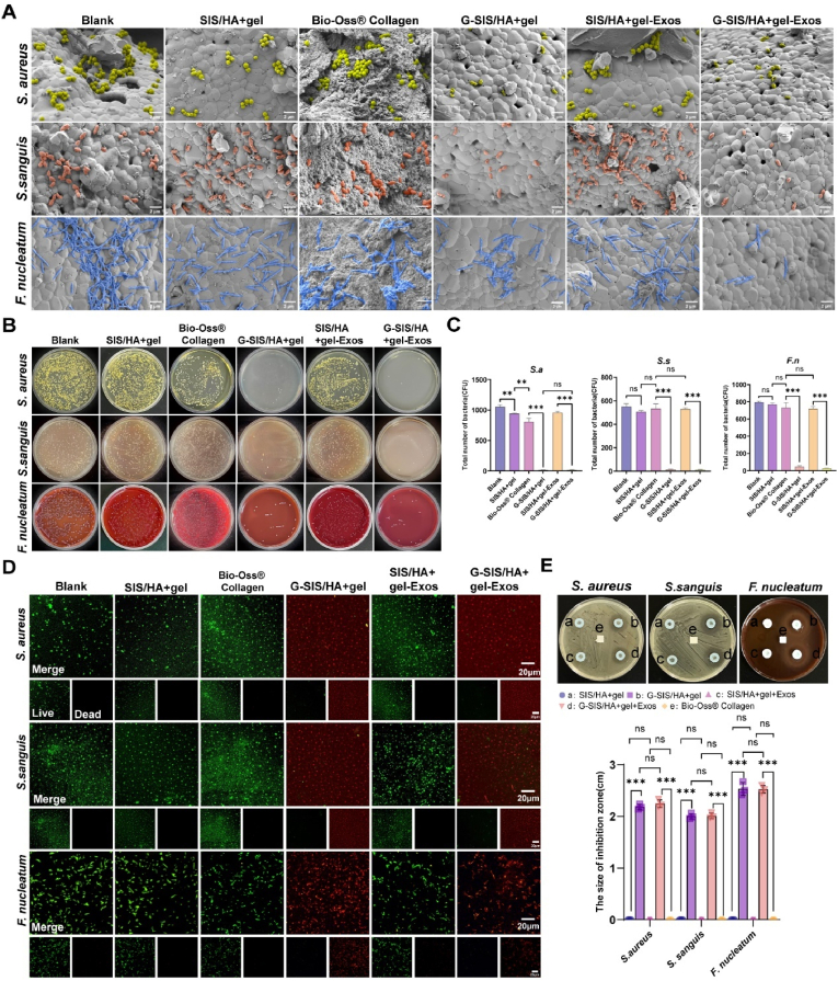
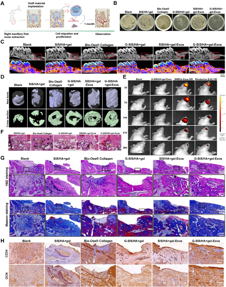
After tooth extraction, alveolar bone absorbs unevenly, leading to soft tissue collapse, which hinders full regeneration. Bone loss makes it harder to do dental implants and repairs. Inspired by the biological architecture of bone, a deformable SIS/HA (Small intestinal submucosa/Hydroxyapatite) composite hydrogel coaxial scaffold was designed to maintain bone volume in the socket. The SIS/HA scaffold containing GL13K as the outer layer, mimicking compact bone, while SIS hydrogel loaded with bone marrow mesenchymal stem cells-derived exosomes (BMSCs-Exos) was utilized as the inner core of the scaffolds, which are like soft tissue in the skeleton. This coaxial scaffold exhibited a modulus of elasticity of 0.82 MPa, enabling it to adaptively fill extraction sockets and maintain an osteogenic space. Concurrently, the inner layer of this composite scaffold, enriched with BMSCs-Exos, promoted the proliferation and migration of human umbilical vein endothelial cells (HUVECs) and BMSCs into the scaffold interior (≈3-fold to the control), up-regulated the expression of genes related to osteogenesis (BMP2, ALP, RUNX2, and OPN) and angiogenesis (HIF-1α and VEGF). This induced new blood vessels and bone growth within the scaffold, addressing the issue of low bone formation rates at the center of defects. GL13K was released by approximately 40.87 ± 4.37 % within the first three days, exerting a localized antibacterial effect and further promoting vascularization and new bone formation in peripheral regions. This design aims to achieve an all-around and efficient bone restoration effect in the extraction socket using coaxial scaffolds through a dual internal and external mechanism.

Keywords: Alveolar ridge preservation; Angiogenesis; Antibacterial; Exosome; Osteogenesis; SIS/HA.


Conflict of interest statement

The authors declare the following personal relationships which may be considered as potential competing interests: Pengfei Wei and Bo Zhao are currently employed by Beijing Biosis Healing Biological Technology Co., Ltd.

Figures

Image 1
Graphical abstract
Fig. 1
Fig. 1
Characterization of BMSCs-derived exosomes. (A) Schematic of exosome synthesis. (B)Representative TEM images of BMSCs-derived Exos. (C) NTA presented the size and particle concentration of BMSCs-Exos. (D) Distribution and intensity of Exos. (E) Western blot analysis of Alix, CD63, CD9, and Cytochrome C expression in BMSCs-derived Exos. (F) CLSM images of SIS hydrogels loaded with BMSCs-Exos. Exos were labeled with DiI. (G) Internalization of DiI-labeled Exos by HUVECs for 24 and 48h. (H)Internalization of DiI-labeled Exos by BMSCs for 24 and 48 h. Nuclei (blue); Exos (red). (I) Release curves of Exos in SIS gel-Exos and SIS/HA + gel-Exos scaffold.
Fig. 2
Fig. 2
Preparation and characterization of coaxial scaffolds. (A) Schematic of coaxial scaffold preparation. (B) Rough appearance of different scaffolds: (a) SIS/HA scaffold (b) Hollow SIS/HA scaffold (c) SIS gel (d) SIS/HA + gel scaffold. (C) SEM images of SIS/HA, SIS gel, SIS/HA + gel, Bio-Oss® Collagen, and G-SIS/HA + gel-Exos scaffolds. (D) Micro CT images of SIS/HA, SIS gel, SIS/HA + gel, Bio-Oss® Collagen, and G-SIS/HA + gel-Exos scaffolds. (E) Images of compression deformation of SIS/HA and SIS/HA + gel scaffolds. (F) Stress-strain curves of different scaffolds. (G) Young's moduli of the different scaffolds. (H) FTIR spectra of SIS/HA, SIS gel, SIS/HA + gel, Bio-Oss® Collagen, and G-SIS/HA + gel-Exos scaffolds. (I) Release profiles of GL13K coaxial scaffold. (J) Degradation curves of SIS/HA, SIS gel, SIS/HA + gel, Bio-Oss® Collagen, and G-SIS/HA + gel-Exos scaffolds.
Fig. 3
Fig. 3
Proliferation, migration, and morphology of cells on different scaffolds. (A) Schematic representation of cell behavior on G-SIS/HA + gel-Exos scaffolds (B) Cell viability of BMSCs and HUVECs at 1, 3, 5, and 7 days (C) Live and dead staining images of BMSCs and HUVECs cultured with different scaffold extracts (1 day). (D) Migration staining of BMSCs and HUVECs with different scaffold extracts. (E) Quantification of the number of migrated BMSCs and HUVECs. One-way analysis of variance was used for statistical analysis. Data are shown as mean ± SD, ∗P <0.05, ∗∗P <0.01, ∗∗∗P <0.001. (F) Scratched wound assay of BMSCs and HUVECs. Quantitative analysis of the area of migration BMSCs and HUVECs. One-way analysis of variance was used for statistical analysis. Data are shown as mean ± SD, ∗P <0.05, ∗∗P <0.01, ∗∗∗P <0.001. Cytoskeletal morphology of (G) BMSCs and (H) HUVECs after 3 days of culture. Nuclei (blue), actin filaments (red).
Fig. 4
Fig. 4
Promotion of angiogenesis in vitro. (A) HUVECs were cultured on Matrigel for 6 h to form microtubules. (B) Expression of angiogenic related proteins (HIF-1α and VEGF) by Western blot analysis. (C) Quantitative analysis of Western blot experiments. Data are shown as mean ± SD, ∗P <0.05, ∗∗P <0.01, ∗∗∗P <0.001. Expression of angiogenesis-related proteins HIF-1α (D) and VEGF (E) was observed by immunofluorescence. (F) Quantitative analysis of fluorescence intensity. Data are shown as mean ± SD, ∗P <0.05, ∗∗P <0.01, ∗∗∗P <0.001. (G) Expression of genes related to angiogenesis (HIF-1α and VEGF). Data are shown as mean ± SD, ∗P <0.05, ∗∗P <0.01, ∗∗∗P <0.001.
Fig. 5
Fig. 5
Studies on osteogenic differentiation in vitro. (A) ALP staining images of different scaffolds at 7 and 14 days. (B) ARS staining images of different scaffolds on 14 and 21 days. (C) Western blot analysis for expression of osteogenesis-related proteins (BMP2, ALP, OPN, and RUNX2) (D) Quantitative analysis of Western blot analysis. Data are shown as mean ± SD, ∗P <0.05, ∗∗P <0.01, ∗∗∗P <0.001. (E) qRT-PCR detection of expression levels of osteogenic-related genes (BMP2, ALP, OPN, and RUNX2). Data are shown as mean ± SD, ∗P <0.05, ∗∗P <0.01, ∗∗∗P <0.001.(F) Immunofluorescence assays of proteins related to osteogenesis.
Fig. 6
Fig. 6
In vitro antibacterial activity (A) SEM image. (B) Plate counts of bacterial colonies (S.aureus, S. sanguis, and F. nucleatum). (C) Quantitative analysis of bacterial colonies. Data are shown as mean ± SD, ∗P <0.05, ∗∗P <0.01, ∗∗∗P <0.001. (D) CLSM images of S. aureus, S. sanguis, and F. nucleatum live/dead staining. Green: live bacteria, red: dead bacteria. (E) Inhibition zones of S. aureus, S. sanguis, and F. nucleatum co-cultured with different scaffolds and statistical analysis of the inhibition zones. (a: SIS/HA + gel, b: G-SIS/HA + gel, c: SIS/HA + gel-Exos, d: G-SIS/HA + gel-Exos, e: Bio-Oss® collagen) Data are shown as mean ± SD, ∗P <0.05, ∗∗P <0.01, ∗∗∗P <0.001.
Fig. 7
Fig. 7
Evaluation and analysis of post-extraction and alveolar reconstruction. (A) Schematic diagram of the experiment. (B) Colony counts at the wound sites. (C) Sagittal picture of the rat maxilla. (D) Three-dimensional reconstructed images of rat alveolar bone defects. (E) G-SIS/HA + gel-Exos materials (Rhodamine B-GL13K, DiR-Exos) were implanted into a rat extraction socket and the release of GL13K and Exos was observed in vivo at different time points. (F) Subcutaneous degradation of different scaffolds in rats (Yellow arrows represent undegraded materials). (G) The newly formed bone tissue was evaluated by H&E staining and Masson's trichrome staining after an operation of 1 month. (H) Immunohistochemical analysis of CD34 and OCN expression.
Fig. 8
Fig. 8
Gene expression and analysis of biological activity of BMSCs cultured in SIS/HA + gel and G-SIS/HA + gel-Exos groups. (A) Venn diagrams and volcano plots of genes affected by coaxial scaffolding. (B) Representative enriched GO up terms of DEGs from SIS/HA + gel versus G-SIS/HA + gel-Exos. (C) Heatmap of DEGs related to cellular activity, angiogenesis and osteogenesis from SIS/HA + gel versus G-SIS/HA + gel-Exos. (D) The enriched KEGG pathways of DEGs from SIS/HA + gel versus G-SIS/HA + gel-Exos. (E) GSEA plots of SIS/HA + gel versus G-SIS/HA + gel-Exos.

Similar articles

References

    1. Aribau-Gumà C., Jorba-García A., Sánchez-Torres A., Sànchez-Garcés M.À. Alveolar ridge preservation: an overview of systematic reviews. Int. J. Oral Maxillofac. Surg. 2022;51:234–242. doi: 10.1016/j.ijom.2021.06.002. - DOI - PubMed
    1. Wang R.E., Lang N.P. Ridge preservation after tooth extraction. Clin. Oral Implants Res. 2012;23:147–156. doi: 10.1111/j.1600-0501.2012.02560.x. - DOI - PubMed
    1. Araújo M.G., Dias D.R., Matarazzo F. Anatomical characteristics of the alveolar process and basal bone that have an effect on socket healing. Periodontology. 2023;93:277–288. doi: 10.1111/prd.12506. 2000. - DOI - PubMed
    1. Chappuis V., Araújo M.G., Buser D. Clinical relevance of dimensional bone and soft tissue alterations post-extraction in esthetic sites. Periodontology. 2017;73:73–83. doi: 10.1111/prd.12167. 2000. - DOI - PubMed
    1. Gu J., Jiao K., Li J., Yan J., Wang K., Wang F., Liu Y., Tay F.R., Chen J., Niu L. Polyphosphate-crosslinked collagen scaffolds for hemostasis and alveolar bone regeneration after tooth extraction. Bioact. Mater. 2022;15:68–81. doi: 10.1016/j.bioactmat.2021.12.019. - DOI - PMC - PubMed

LinkOut - more resources