. 2025 Apr;9(4):e2400538.
doi: 10.1002/adbi.202400538. Epub 2025 Jan 6.

Depletion of TP53 in Human Pluripotent Stem Cells Triggers Malignant-Like Behavior

Affiliations

Depletion of TP53 in Human Pluripotent Stem Cells Triggers Malignant-Like Behavior

Joaquin Montilla-Rojo et al. Adv Biol (Weinh). 2025 Apr.

Abstract

Human pluripotent stem cells (hPSCs) tend to acquire genetic aberrations upon culture in vitro. Common aberrations are mutations in the tumor suppressor TP53, suspected to confer a growth-advantage to the mutant cells. However, their full impact in the development of malignant features and safety of hPSCs for downstream applications is yet to be elucidated. Here, TP53 is knocked out in hPSCs using CRISPR-Cas9 and compared them with isogenic wild-type hPSCs and human germ cell tumor lines as models of malignancy. While no major changes in proliferation, pluripotency, and transcriptomic profiles are found, mutant lines display aberrations in some of the main chromosomal hotspots for genetic abnormalities in hPSCs. Additionally, enhanced clonogenic and anchorage-free growth, alongside resistance to chemotherapeutic compounds is observed. The results indicate that common TP53-depleting mutations in hPSCs, although potentially overlooked by standard analyses, can impact their behavior and safety in a clinical setting.

Keywords: TP53; genetic aberration; human germ cell tumor; human pluripotent stem cell; malignancy; pluripotency.


Conflict of interest statement

The authors declare no conflict of interest.

Figures

Figure 1
Figure 1
Generation of TP53 knockout hPSC lines. A) SnapGene genome sequence alignments of the CRISPR/Cas9 target site of the TP53 gene in H9‐KO as a representative of the knockout methodology. The knockout cell line (bottom sequence) shows a one‐base‐pair insertion (A, adenine), resulting in a premature STOP codon. B) Representative images of colony morphology for all hPSC lines and generated isogenic TP53‐knockout clones. Scalebars represent 200 µm C) RT‐qPCR data and western blot showing the loss of TP53 both in mRNA and protein levels, respectively. D) Flow cytometric data showing the (lack of) differences in expression of key pluripotency markers between the different hPSC lines and their respective isogenic TP53‐depleted counterparts. E) Bar graph showing the fold change in gene expression, as determined by RT‐qPCR, between parental cell lines and their isogenic TP53‐knockout clones. Graphs represent results from at least three independent experiments (n≥3). Bars indicate mean; error bars indicate standard deviation. p‐values were calculated using unpaired Student's t‐test. *p ≤ 0.05.
Figure 2
Figure 2
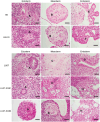
Assessment of differentiation capacity in embryoid bodies. Representative sections of H&E‐stained embryoid bodies showing differentiated structures derived from the three germ layers: A) Neural rosette; B) Cartilage; C) Bone; D) Hematopoietic precursors; E) Intestinal‐like epithelium; F) Glandular tissue; G) Ciliated lung epithelium; H) Adipose tissue. Scale bars represent 50 µm.
Figure 3
Figure 3
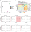
Transcriptomic and genetic stability analysis of wild‐type and TP53‐knockout hPSC and hGCT lines. A) Principal Component Analysis of mRNA expression of wild‐type and TP53‐knockout hPSC and hGCT lines. B) Hierarchical clustering using a TP53 target gene set showing the resemblance of hPSCs and hGCTs as well as confirming the effects of depleting TP53 in the transcriptome of the different lines. C) GSA profiling of LU07, LU07‐KO#1, and LU07‐KO#2, showing the presence of copy number amplifications in chromosomes 16q and 20q11 for the TP53‐knockout clones compared to their respective wild‐type isogenic line. Areas highlighted in red represent amplified regions.
Figure 4
Figure 4
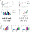
Phenotypic characterization of TP53 wild‐type and knockout hPSC and hGCTs. A) Cell proliferation rate comparison for hPSC (left) and hGCT lines (right) with their respective TP53 knockout isogenic counterparts. B) Clonogenic capacity of the different lines upon low‐density seeding as measured on the basis of total area per well covered by cell clusters and cell density per cluster. Representative pictures of one biological replicate are displayed. C. Soft‐agar assay, number of colonies grown upon seeding of the different lines in medium containing low‐concentration agarose. Representative pictures of one biological replicate are displayed. D) Bar graphs showing the viability of wild‐type cell lines and their corresponding knockout clones when treated with cisplatin, gemcitabine, and navitoclax for 72 h. All graphs represent results from at least three independent experiments (n ≥ 3). 21‐K and NC‐K stand for 2102Ep‐KO and NCCIT‐KO, respectively. Cisplatin IC50 data for 2102Ep and NCCIT TP53‐WT and ‐KO lines, represented by bars highlighted with a diagonal dotted line in panel D, have been taken from previous experiments published elsewhere,[ 39 ] to display a better overview of the impact of these mutations in the resistance to cisplatin regardless of the cell type. Data presented as mean; error bars represent standard deviation. p‐values were calculated using unpaired Student's t‐test (Panels B and C) or one‐way ANOVA with Tukey's multiple comparisons post hoc test (Panels A and D). *p ≤ 0.05; **p ≤ 0.01; # p ≤ 0.0001; ns = not significant.

Similar articles

Cited by

References

    1. Sguazzi G. P., Muto V., Tartaglia M., Bertini E., Compagnucci C., Int. J. Mol. Sci. 2021, 22, 13674. - PMC - PubMed
    1. Madrid M., Sumen C., Aivio S., Saklayen N., Curr. Protoc. 2021, 1, e88. - PubMed
    1. Chehelgerdi M., Behdarvand Dehkordi F., Chehelgerdi M., Kabiri H., Salehian‐Dehkordi H., Abdolvand M., Salmanizadeh S., Rashidi M., Niazmand A., Ahmadi S., Feizbakhshan S., Kabiri S., Vatandoost N., Ranjbarnejad T., Mol. Cancer 2023, 22, 189. - PMC - PubMed
    1. Merkle F. T., Ghosh S., Kamitaki N., Mitchell J., Avior Y., Mello C., Kashin S., Mekhoubad S., Ilic D., Charlton M., Saphier G., Handsaker R. E., Genovese G., Bar S., Benvenisty N., McCarroll S. A., Eggan K., Nature 2017, 545, 229. - PMC - PubMed
    1. Lezmi E., Benvenisty N., Nat. Protoc. 2021, 16, 4522. - PubMed

MeSH terms

Substances

LinkOut - more resources