

Rhus Verniciflua Stokes Inhibits PD-1 Expression and Induces Anticancer Effects by Enhancing T Cell Function
- PMID: 39760460
- PMCID: PMC11705362
- DOI: 10.1177/15347354241308220
Rhus Verniciflua Stokes Inhibits PD-1 Expression and Induces Anticancer Effects by Enhancing T Cell Function
Abstract
Background: Over the last decade, the anticancer effects of Rhus verniciflua Stokes (RVS) have been reported in various preclinical or clinical studies. However, the effects of RVS on immuno-oncology, especially on the functional properties of T cells and their phenotypes, remain unclear. Here, we planned to investigate the impact of RVS on immuno-oncology, specifically focusing on its effects on T cells.
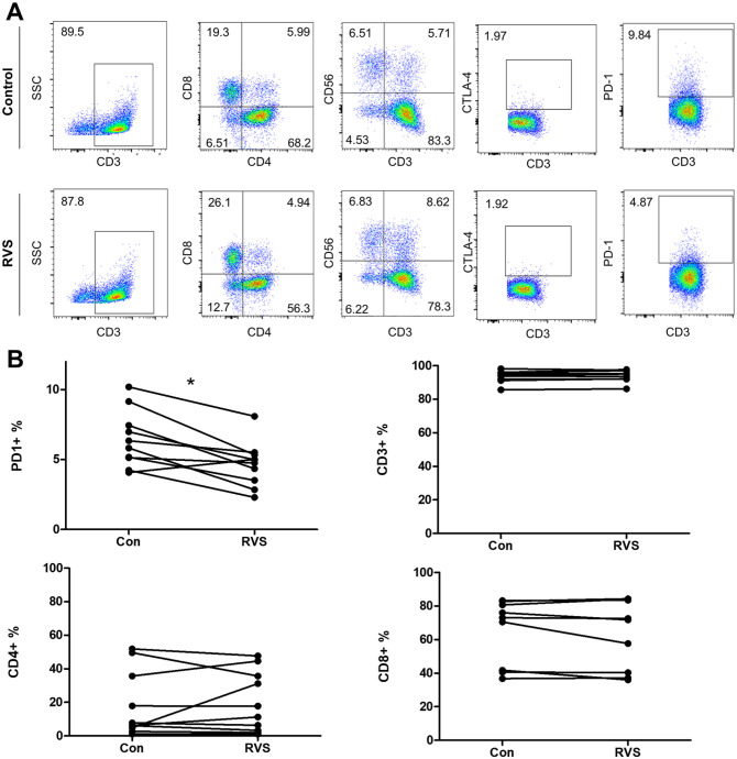
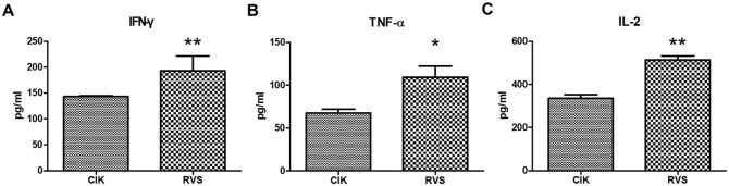
Methods: Peripheral blood mononuclear cells (PBMCs) from breast cancer patients were isolated to obtain cytokine-induced killer cell populations with >85% CD3+ T cells. The anticancer activity of these T cells was evaluated by introducing red fluorescent protein (RFP) into HLA-A02:01 type-matched breast cancer cell lines (MCF7 and MDA-MB-231) and analyzing the results using flow cytometry. The effect of RVS extracts on T cell phenotype was assessed using markers such as CTLA-4 and PD-1, as well as mRNA expression levels of key genes (IFN-γ, TNF-α, and IL-2).
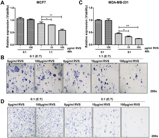
Results: RVS treatment significantly enhanced the anticancer activity of T cells against breast cancer cells. Specifically, T cells treated with 100 µg/mL of RVS showed a 20.6% increase in cytotoxicity against MCF-7 cells and a 36.2% increase against MDA-MB231 cells compared to the control. Additionally, RVS treatment led to a significant reduction in PD-1 expression on T cells.
Conclusion: Our findings demonstrate that RVS treatment enhances T cell function against breast cancer cells by reducing PD-1 expression. These results suggest that components of RVS may serve as potential candidates for restoring exhausted T cells in cancer therapy.
Keywords: PD-1; Rhus verniciflua Stokes; T cell co-culture; anticancer; breast cancer; immuno-oncology.
Conflict of interest statement
Declaration of Conflicting InterestsThe author(s) declared no potential conflicts of interest with respect to the research, authorship, and/or publication of this article.
Figures
References
-
- Li MC, Zhang Y-Q, Meng C-W, et al.. Traditional uses, phytochemistry, and pharmacology of Toxicodendron vernicifluum (Stokes) F.A. Barkley - a review. J Ethnopharmacol. 2021;267:113476. - PubMed
-
- Lee SH, Kim KS, Choi WC, Yoon SW. The concurrent use of rhus verniciflua stokes as complementary therapy with second or more line regimens on advanced non-small-cell lung cancer: case series. J Korean Med. 2009;30:112-117.
-
- Lee SH, Choi WC, Yoon SW. Impact of standardized Rhus verniciflua stokes extract as complementary therapy on metastatic colorectal cancer: a Korean single-center experience. Integr Cancer Ther. 2009;8:148-152. - PubMed
-
- Lee SK, Jung HS, Eo WK, et al.. Rhus verniciflua Stokes extract as a potential option for treatment of metastatic renal cell carcinoma: report of two cases. Ann Oncol. 2010;21:1383-1385. - PubMed
Publication types
MeSH terms
Substances
LinkOut - more resources
Full Text Sources
Medical
Research Materials
Miscellaneous
