. 2025 Jan 6;40(1):89.
doi: 10.1007/s11011-024-01513-6.

Metabolomics analyses and comparative insight to neuroprotective potential of unripe fruits and leaves of Citrus aurantium ethanolic extracts against cadmium-induced rat brain dysfunction: involvement of oxidative stress and akt-mediated CREB/BDNF and GSK3β/NF-κB signaling pathways

Affiliations

Metabolomics analyses and comparative insight to neuroprotective potential of unripe fruits and leaves of Citrus aurantium ethanolic extracts against cadmium-induced rat brain dysfunction: involvement of oxidative stress and akt-mediated CREB/BDNF and GSK3β/NF-κB signaling pathways

Doaa A H Deabes et al. Metab Brain Dis. .

Abstract

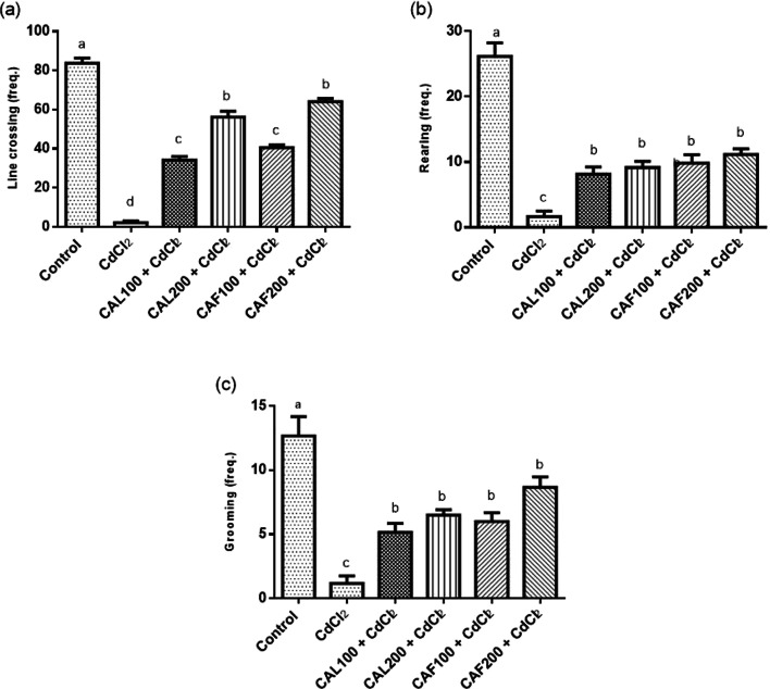
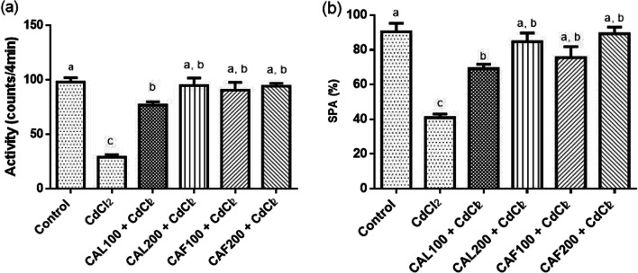
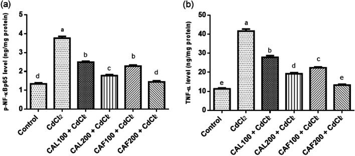
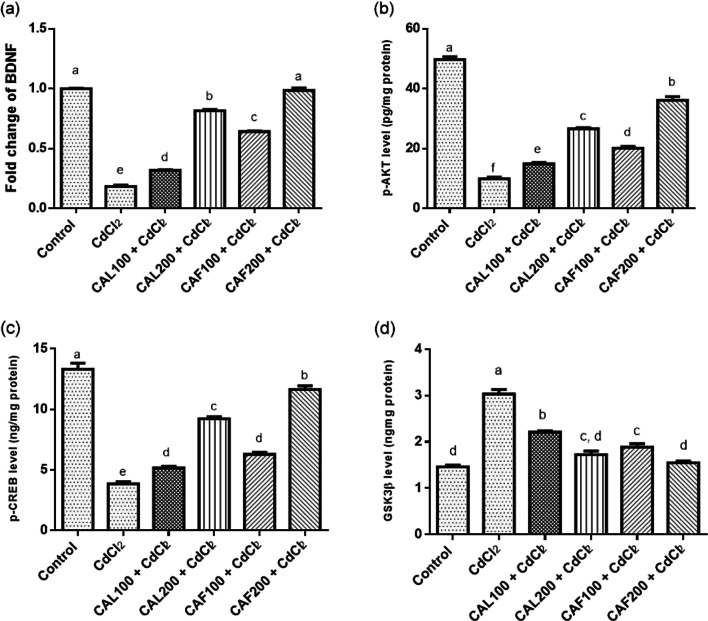
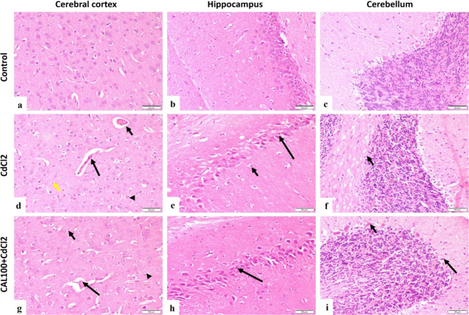
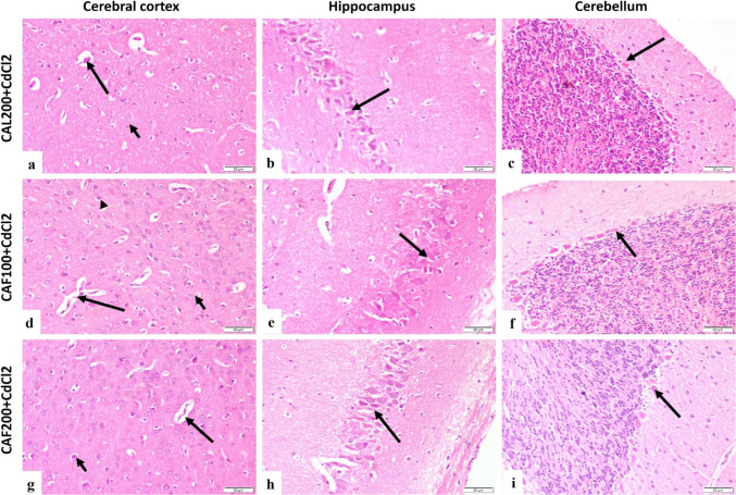
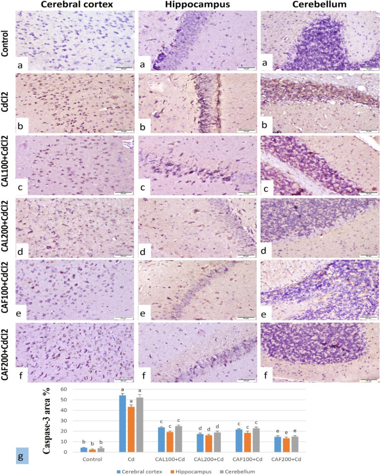
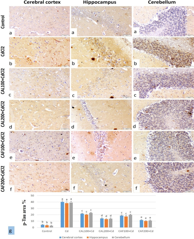
Serious neurological disorders were associated with cadmium toxicity. Hence, this research aimed to investigate the potential neuroprotective impacts of the ethanolic extracts of Citrus aurantium unripe fruits and leaves (CAF and CAL, respectively) at doses 100 and 200 mg/kg against cadmium chloride-provoked brain dysfunction in rats for 30 consecutive days. HPLC for natural pigment content revealed that CAF implied higher contents of Chlorophyll B, while the CAL has a high yield of chlorophyll A and total carotenoid. Fifty-seven chromatographic peaks were identified by UPLC/MS/MS; 49 and 29 were recognized from CAF or CAL, respectively. Four compounds were isolated from CAF: 3',4',7 -trihydroxyflavone, isorhainetin, vitexin, and apigenin. In vitro studies outlined the antioxidant capacity of studied extracts where CAF showed better scavenging radical DPPH activity. Results clarified that both extracts with a superior function of CAF at the high adopted dose significantly ameliorated CdCl2-induced neuro-oxidative stress and neuro-inflammatory response via restoring antioxidant status and hindering nuclear factor kappa B (NF-κB) stimulation. Moreover, it up-regulated the levels of phospho-protein kinase B (p-Akt), phospho- cAMP-response element binding protein (p-CREB), and brain-derived neurotropic factor (BDNF) levels, and elicited a marked decrease in the content of glycogen synthase kinase 3 beta (GSK3β), besides amending Caspase-3 and hyperphosphorylation of tau protein in brain tissues. Moreover, a significant improvement in the rats' behavioral tasks of the CAL and CAF-treated groups has been recorded, as indicated by marked preservation in locomotion, exploratory, and memory functions of the experimental rats. In conclusion, the reported neuroprotective impacts of C. aurantium extracts may be through modulating p-AKT/p-CREB/BDNF and / or p-Akt/ GSK3β/NF-κB signaling pathways.

Keywords: Citrus aurantium; Cadmium; Cognitive function; Flavonoids; Neurotoxicity.


Conflict of interest statement

Declarations. Ethical approval: This work was carried out in compliance with the ethical procedures and policies delineated in the National Institutes of Health guide for the care and use of Laboratory animals (NIH Publications No. 8023, revised 1978)., the ARRIVE guidelines and international legislation, which approved by the Medical Research Ethics (MREC) Committee of the National Research Centre, Dokki, Giza, Egypt (Approval no. 7441201-2023). Consent to participate: Not applicable. Consent to Publish: Not applicable. Competing interests: The authors declare no competing interests.

Figures

Fig. 1
Fig. 1
Ameliorative effects of C. aurantium extracts on rats’ behavioral changes in the open field task. Where, (a) line crossing, (b) rearing and (c) grooming frequencies. Each bar represents the mean ± SE (n = 6) of individual group and the significance at p < 0.05 is illustrated by distinct letters using Tukey’s post-hoc test
Fig. 2
Fig. 2
Ameliorative effects of C. aurantium extracts on the changes of activity cage and Y maze. Where, (a) activity counts and (b) SPA% recorded in activity cage and Y maze, respectively. Each bar represents the mean ± SE (n = 6) of individual group and the significance at p < 0.05 is illustrated by distinct letters using Tukey’s post-hoc test
Fig. 3
Fig. 3
Ameliorative effects of C. aurantium extracts on the changes of inflammatory markers in the brain. Where, (a) pNF-κBp65 and (b) TNF-α. Each bar represents the mean ± SE (n = 6) of individual group and the significance at p < 0.05 is illustrated by distinct letters using Tukey’s post-hoc test
Fig. 4
Fig. 4
Ameliorative effects of C. aurantium extracts on changes of brain p-Akt/p-CREB/BDNF/GSK3β signaling pathway. Where, (a) BDNF gene expression, (b) p-AKT, (c) p-CREB and (d) GSK3β. Each bar represents the mean ± SE (n = 6) of individual group and the significance at p < 0.05 is illustrated by distinct letters using Tukey’s post-hoc test
Fig. 5
Fig. 5
Photomicrograph, rat brain, (H&E, scale bar 50 μm). (a-c) cerebral cortex, hippocampus and cerebellum of control group showing normal histological structure. (d) CdCl2 group cerebral cortex showing capillary congestion (short arrow), capillary endothelial proliferation (long arrow), neuronal degeneration with neuronophagia (arrowhead) and astrocytosis (yellow arrow). (e) CdCl2 group hippocampus showing pyramidal neurons degeneration (long arrow) and astrocytosis (short arrow). (f) CdCl2 group cerebellum showing degeneration of purkinje cells (arrow). (g) CAL100 + CdCl2 group cerebral cortex showing moderately congested cerebral capillary (long arrow), astrocytosis (short arrow) and few degenerated neurons (arrowhead). (h) CAL100 + CdCl2 group hippocampus showing few degenerated pyramidal neurons (arrow). (i) CAL100 + CdCl2 group cerebellum showing degenerated purkinje cells (long arrow) and normal purkinje cells (short arrow)
Fig. 6
Fig. 6
Photomicrograph, rat brain, (H&E, scale bar 50 μm). (a) CAL200 + CdCl2 group cerebral cortex showing moderately congested capillary (long arrow) and astrocytosis (short arrow). (b) CAL200 + CdCl2 group hippocampus showing nearly normal neurons (arrow). (c) CAL200 + CdCl2 group cerebellum showing normal purkinje cells (arrow). (d) CAF100 + CdCl2 group cerebral cortex showing mild capillary proliferation (long arrow), astrocytosis (short arrow) and neuronal degeneration (arrowhead). (e) CAF100 + CdCl2 group hippocampus showing few degenerated neurons (arrow). (f) CAF100 + CdCl2 group cerebellum showing nearly normal purkinje cells (arrow). (g) CAF200 + CdCl2 group cerebral cortex showing normal cerebral capillary (long arrow) and few degenerated neurons (short arrow). (h) CAF200 + CdCl2 group hippocampus showing normal neurons (arrow). (i) CAF200 + CdCl2 group cerebellum showing normal purkinje cells (arrow)
Fig. 7
Fig. 7
Immunostaining of caspase-3 of rat brain (Caspase-3, scale bar 50 μm). (a) Cerebral cortex, hippocampus and cerebellum of control group showing very weak to nil immuno-reaction. (b) CdCl2 group cerebral cortex, hippocampus and cerebellum showing strong immune-expression. (c) CAL100 + CdCl2 group showing moderate to weak positive immuno-reaction. (d) CAL200 + CdCl2 group showing weak immune-reactive cells. (e) CAF100 + CdCl2 group showing moderate to weak immune-expression. (f) CAF200 + CdCl2 group showing weak caspase-3 expression. (g) caspase-3 expression % area in brain tissue of different treated groups (Data was expressed as mean ± SE, dissimilar letters designating significant differences at p < 0.05)
Fig. 8
Fig. 8
Immunostaining of p-Tau protein of rat brain (p-Tau, scale bar 50 μm). (a) Cerebral cortex, hippocampus and cerebellum of control group showing very weak to no immuno-reactive cells. (b) CdCl2 group showing strong immune-expression in cerebral cortex, hippocampus and cerebellum. (c) CAL100 + CdCl2 group showing moderate positive immuno-reaction. (d) CAL200 + CdCl2 group showing moderate to weak expression. (e) CAF100 + CdCl2 group showing moderate immune-expression (f) CAF200 + CdCl2. group showing weak expression. (g) p-Tau expression % area in brain tissue of different treated groups (Data was expressed as mean ± SE, dissimilar letters designating significant differences at p < 0.05)

Similar articles

References

    1. Abdel-Aleem GA, Khaleel EF (2018) Rutin hydrate ameliorates cadmium chloride-induced spatial memory loss and neural apoptosis in rats by enhancing levels of acetylcholine, inhibiting JNK and ERK1/2 activation and activating mTOR signalling. Arch Physiol Biochem 124:367–377 - PubMed
    1. Afifi OK, Embaby AS (2016) Histological study on the protective role of ascorbic acid on cadmium induced cerebral cortical neurotoxicity in adult male albino rats. J Microscopy Ultrastructure 4:36–45 - PMC - PubMed
    1. Al Olayan EM, Aloufi AS, AlAmri OD, Ola H, Moneim AEA (2020) Protocatechuic acid mitigates cadmium-induced neurotoxicity in rats: role of oxidative stress, inflammation and apoptosis. Sci Total Environ 723:137969 - PubMed
    1. Al-Majedy YK, Al-Duhaidahawi DL, Al-Azawi KF, Al-Amiery AA, Kadhum AAH, Mohamad AB (2016) Coumarins as potential antioxidant agents complemented with suggested mechanisms and approved by molecular modeling studies. Molecules 21:135 - PMC - PubMed
    1. Alqudah A, Qnais EY, Wedyan MA, Altaber S, Bseiso Y, Oqal M, AbuDalo R, Alrosan K, Alrosan AZ, Bani Melhim S (2023) Isorhamnetin reduces glucose level, inflammation, and oxidative stress in high-fat diet/Streptozotocin diabetic mice model. Molecules 28:502 - PMC - PubMed

MeSH terms

LinkOut - more resources