. 2025 Jan 6;21(1):e1012787.
doi: 10.1371/journal.ppat.1012787. eCollection 2025 Jan.

Isoferulic acid facilitates effective clearance of hypervirulent Klebsiella pneumoniae through targeting capsule

Affiliations

Isoferulic acid facilitates effective clearance of hypervirulent Klebsiella pneumoniae through targeting capsule

Tingting Wang et al. PLoS Pathog. .

Abstract

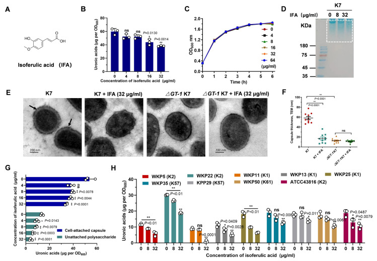
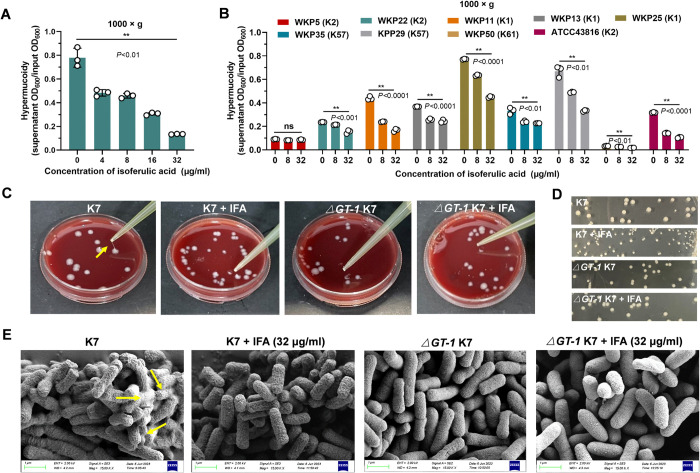
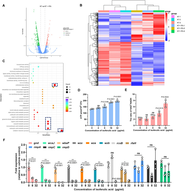
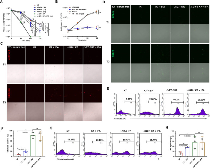
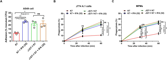
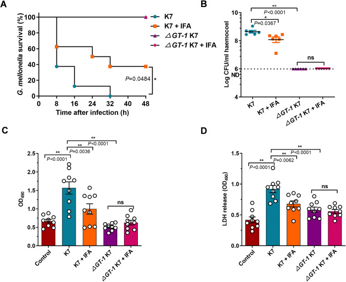
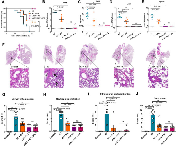
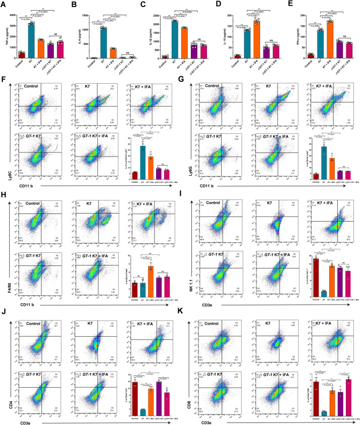
Hypervirulent Klebsiella pneumoniae (hvKP) poses an alarming threat in clinical settings and global public health owing to its high pathogenicity, epidemic success and rapid development of drug resistance, especially the emergence of carbapenem-resistant lineages (CR-hvKP). With the decline of the "last resort" antibiotic class and the decreasing efficacy of first-line antibiotics, innovative alternative therapeutics are urgently needed. Capsule, an essential virulence determinant, is a major cause of the enhanced pathogenicity of hvKP and thus represents an attractive drug target to prevent the devastating clinical outcomes caused by hvKP infection. Here, we identified isoferulic acid (IFA), a natural phenolic acid compound widely present in traditional herbal medicines, as a potent broad-spectrum K. pneumoniae capsule inhibitor that suppresses capsule polysaccharide synthesis by increasing the energy status of bacteria. In this way, IFA remarkably reduced capsule thickness and impaired hypercapsule-associated hypermucoviscosity phenotype (HMV), thereby significantly sensitizing hvKP to complement-mediated bacterial killing and accelerating host cell adhesion and phagocytosis. Consequently, IFA facilitated effective bacterial clearance and thus remarkably protected mice from lethal hvKP infection, as evidenced by limited bacterial dissemination and a significant improvement in survival rate. In conclusion, this work promotes the development of a capsule-targeted alternative therapeutic strategy for the use of the promising candidate IFA as an intervention to curb hvKP infection, particularly drug-resistant cases.


Conflict of interest statement

The authors have declared that no competing interests exist.

Figures

Fig 1
Fig 1. IFA effectively reduced the production of K. pneumoniae capsule.
(A) Chemical structure of IFA (IFA). (B) Uronic acid assay to confirm the capsule production of K. pneumoniae K7 with the treatment of indicated concentrations of IFA. (C) Growth curves of K. pneumoniae K7 in the presence of the indicated concentrations of IFA. (D) Alcian blue staining of capsule samples extracted from K. pneumoniae K7 with or without IFA treatment. (E) Transmission electron microscopy images of wild-type K. pneumoniae K7 and ΔGT-1 K7 in the presence of DMSO or 32 μg/ml IFA. The black arrows indicate capsule, and its thickness was measured using ImageJ (F). (G) Comparison of cell-attached and unattached capsule content of K. pneumoniae K7 with or without IFA treatment via a uronic acid assay. (H) Capsule production of different K. pneumoniae strains with or without IFA treatment. The data are presented as the means ± SEMs. One-way ANOVA and Tukey’s posttest was carried out to determine the statistical significance of different groups. *P < 0.05, **P < 0.01 and ns, no significance compared with the DMSO control group.
Fig 2
Fig 2. IFA abrogated capsule-associated hypermucoviscosity phenotypes of hypervirulent K. pneumoniae.
(A) Hypermucoidy assay of hypervirulent K. pneumoniae K7 treated with DMSO or indicated concentrations of IFA. Bacterial cultures were centrifuged at 1,000 × g in a fixed angle rotor for 5 min to measure the OD600 of the supernatants, which was defined relative to the initial OD600. (B) Hypermucoidy analysis of different serotypes of K. pneumoniae. (C) Representative colony phenotypes of K. pneumoniae K7 and ΔGT-1 K7 on blood agar plates containing DMSO or 32 μg/ml IFA; the “string test” was performed at the same time. The yellow arrow indicates the string stretched by the tips. (D) The colony morphologies of K. pneumoniae K7 and ΔGT-1 K7 on LB agar plates containing DMSO or 32 μg/ml IFA are shown. (E) Scanning electron microscopy images of wild-type K. pneumoniae K7 and ΔGT-1 K7 in the presence of DMSO or 32 μg/ml IFA. The data are presented as the means ± SEMs. One-way ANOVA and Tukey’s posttest was performed to determine the statistical significance of different groups. **P < 0.01 and ns, no significance compared with the DMSO control group.
Fig 3
Fig 3. IFA reduced the capsular biosynthesis through altering bacterial metabolism.
(A) Volcano plot analysis of differentially expressed genes (DEGs) between DMSO- or 32 μg/ml IFA-treated K. pneumoniae K7. A total of 295 differentially expressed genes (DEGs), including 78 upregulated and 217 downregulated genes, were identified in the IFA-treated group. (B) Heat cluster of DEGs. (C) The top 20 enriched Gene Ontology (GO) terms of all DEGs compared to DMSO control. (D) Intracellular ATP levels in K. pneumoniae K7 cells treated with DMSO or indicated concentrations of IFA. (E) The ratio of NAD+/NADH in K. pneumoniae K7 treated with DMSO or indicated concentrations of IFA. (F) The transcriptional levels of capsule synthesis genes and rmp genes. The data are presented as the means ± SEMs. One-way ANOVA and Tukey’s posttest was performed to determine the statistical significance of different groups.*P < 0.05, **P < 0.01 and ns, no significance compared with DMSO control group.
Fig 4
Fig 4. IFA sensitized hypervirulent K. pneumoniae to complement-mediated killing by inhibiting capsule.
(A) Serum-killing assay of K. pneumoniae K7 and ΔGT-1 K7 in the presence of DMSO or indicated concentrations of IFA. Bacteria were incubated with 20% NHS, and the viable count was determined by serial dilution and microbiological plating at the indicated time points. (B) Bacterial viability of K. pneumoniae K7 in heat-inactivated NHS (HNHS) and LB medium supplemented with DMSO or 64 μg/ml IFA. (C) Immunofluorescence microscopy analysis of C3b/C3bi deposition on the surface of 1-hour and 3-hour serum-exposed K. pneumoniae K7 and ΔGT-1 K7 with the treatment of DMSO or 32 μg/ml IFA. C3b/C3bi was stained with APC-conjugated anti-C3b/C3bi antibody. (D) Immunofluorescence microscopy analysis of C5b-9 formation on the surface of 1-hour- and 3-hour serum-exposed K. pneumoniae K7 and ΔGT-1 K7 with the treatment of DMSO or 32 μg/ml IFA. The C5b-9 complex was stained with a mouse anti-C5b-9 antibody and Alexa Fluor 488-conjugated goat anti-mouse IgG. The fluorescence images (top) and transillumination images (bottom) were normalized within each panel. (E) Flow cytometry-based determination of C3b/C3bi binding to K. pneumoniae K7 and ΔGT-1 K7 with the treatment of DMSO or 32 μg/ml IFA was performed after a 3-hour incubation in 20% NHS at 37°C. (F) The data were analyzed using unpaired two-tailed Student’s t-test. (n = 3). (G) Flow cytometry-based determination of C5b-9 formation on K. pneumoniae K7 and ΔGT-1 K7 with the treatment of DMSO or 32 μg/ml IFA was performed after 3 hours of incubation in 20% NHS at 37°C. (H) The data were analyzed using unpaired two-tailed Student’s t-test (n = 3). The means ± SEMs are shown. *P < 0.05, **P < 0.01 and ns, no significance.
Fig 5
Fig 5. IFA disarmed capsule-mediated immune evasion.
(A) Adhesion assay of K. pneumoniae K7 and ΔGT-1 K7 to A549 cells in the presence of DMSO or 32 μg/ml IFA. Cells were infected with K. pneumoniae K7 or ΔGT-1 K7 at an MOI of 50 for 2 hours and then washed, lysed and plated on LB agar plates to quantify the number of colony-forming units. Data are presented as a percentage of the initial inoculum CFU. (B) Phagocytosis of K. pneumoniae K7 and ΔGT-1 K7 by J774 macrophages in the presence of DMSO or 32 μg/ml IFA. The cells were infected at an MOI of 5 for 2 hours and then washed, followed by a further incubation in medium containing gentamicin (100 μg/ml) to kill the extracellular bacteria. The cells were then rinsed, lysed and plated on LB agar plates after serial dilution. (C) Phagocytosis of K. pneumoniae K7 and ΔGT-1 K7 by mouse primary peritoneal macrophages (MPMs) in the presence of DMSO or 32 μg/ml IFA was also analyzed as described above. The data are presented as the means ± SEMs. Unpaired two-tailed Student’s t-test was performed to determine the statistical significance of two groups. *P < 0.05, **P < 0.01 and ns, no significance.
Fig 6
Fig 6. IFA reduced the virulence of hypervirulent K. pneumoniae in G. mellonella larvae.
(A) Survival of G. mellonella 48 h after K. pneumoniae infection. G. mellonella were treated with 50 mg/kg IFA in 10 μl of 10% DMSO containing vehicle (10% DMSO, 45% stroke-physiological saline solution, 40% PEG400 and 5% Tween-80) or an equal volume of vehicle immediately after challenge with 2 × 10⁴ wild-type K. pneumoniae K7 or ΔGT-1 K7 bacteria, and the number of deaths was recorded for survival analysis. Statistical analysis was performed using the log-rank (Mantel–Cox) test (n = 8 larvae each group). (B) Bacterial load in the hemocoels of larvae (presented as cfu/ml). G. mellonella infected and treated as described above were sacrificed at 5 hours postinfection, and the hemocoels were serially diluted and microbiologically plated (n = 6 larvae each group). ND, not detected. The back lines presents the means ± SEMs. (C) Infection-induced melanization of hemocoels from the indicated groups. G. mellonella infected and treated as described above were sacrificed at 5 hours postinfection, and the hemocoels were collected to measure the degree of melanization at OD490nm (n = 9 larvae each group). The larvae in control group were injected with PBS and treated by equal volume of vehicle. (D) LDH release in the hemocoel from the indicated groups. G. mellonella infected as described above were treated with 50 mg/kg IFA in 10 μl of 1% DMSO containing vehicle (1% DMSO, 54% stroke-physiological saline solution, 40% PEG400 and 5% Tween-80) or an equal volume of vehicle and were sacrificed at 5 hours postinfection. Subsequently, the hemocoel was collected to measure LDH release using a cytotoxicity detection kit (n = 9 larvae each group). The results are presented as the OD490nm values. The data are presented as the means ± SEMs. One-way ANOVA and Tukey’s posttest was used to perform multiple comparisons. *P < 0.05, **P < 0.01 and ns, no significance.
Fig 7
Fig 7. IFA effectively limited bacterial dissemination and protected mice from lethal hypervirulent K. pneumoniae pneumonia.
(A) Survival of mice 72 hours after K. pneumoniae infection. Mice were subcutaneously injected with 50 mg/kg IFA in 50 μl of 10% DMSO containing vehicle (10% DMSO, 45% stroke-physiological saline solution, 40% PEG400 and 5% Tween-80) or an equal volume of vehicle immediately after challenge with 1 × 10⁷ wild-type K. pneumoniae K7 or ΔGT-1 K7 bacteria, and the number of deaths was recorded for survival analysis. Statistical analysis was performed using the log-rank (Mantel–Cox) test (n = 12 mice each group). (B) Bacterial burden in the lung tissues of sublethally infected mice. Mice challenged with 2.5 × 10⁶ wild-type K. pneumoniae K7 or ΔGT-1 K7 bacteria were treated as described above and sacrificed at 40 hours post infection, and lung tissues were removed and homogenized in sterilized PBS (10% w/v) to analyze the bacterial burden by microbiological plating (n = 5 mice each group). (C) Bacterial load in the bronchoalveolar lavage fluid (BALF) obtained from sublethally infected mice. Mice that were infected and treated as described above were sacrificed at 32 hours post infection for bronchoalveolar lavage (BAL) analysis, and the BALF was serially diluted and microbiologically plated (n = 6 mice in each group). The bacterial loads in the livers (D) and spleens (E) from the mice mentioned in (B) were also evaluated by microbiologically plating the tissue homogenates in PBS (n = 5 mice each group). ND, not detected. The back lines present the means ± SEMs. (F) H&E-stained lung tissues from mice infected and treated as indicated. The arrows indicate neutrophil infiltration. The degree of airway inflammation (G), neutrophilic infiltration (H), intralesional bacterial burden (I) and total histopathology score (J) were assessed according to standard pathology criteria by blinded scoring. The data are presented as the means ± SEMs. Unpaired two-tailed Student’s t-test was performed to determine the statistical significance of two groups. *P < 0.05, **P < 0.01 and ns, no significance.
Fig 8
Fig 8. IFA treatment facilitated host immune clearance of hypervirulent K. pneumoniae by targeting capsule.
Mice challenged with 2.5 × 10⁶ wild-type K. pneumoniae K7 or ΔGT-1 K7 bacteria were subcutaneously injected with 50 mg/kg IFA in 50 μl of 10% DMSO containing vehicle (10% DMSO, 45% stroke-physiological saline solution, 40% PEG400 and 5% Tween-80) or an equal volume of vehicle, and were then sacrificed at 40 hours postinfection. The concentrations of TNF-α(A), IL-6 (B), IL-1β(C), IL-10 (D) and IFN-γ (E) in lung tissues were measured using ELISA kits following the manufacturer’s instructions (n = 5 mice each group). To determine the number of immune cells in the BALF, mice infected and treated as indicated were sacrificed at 32 h postinfection for BALF collection. The populations of inflammatory monocytes (CD11b+Ly6C+) (F), neutrophils (CD11b+Ly6G+) (G), mature macrophages (CD11b+F4/80+) (H), NK cells (CD3e+NK1.1+) (I), CD4+ T cells (CD3e+CD4+) (J) and CD8+ T cells (CD3e+CD8+) (K) in the BALF from the indicated groups were quantified by flow cytometry (n = 3 mice each group). The y-axis in F-K means the percentage of gated 30000 cells from individual mice BALF for each marker set. The data are presented as the means ± SEMs. Unpaired two-tailed Student’s t-test was performed to determine the statistical significance of two groups. *P < 0.05, **P < 0.01 and ns, no significance.

Similar articles

References

    1. Diekema DJ, Hsueh PR, Mendes RE, Pfaller MA, Rolston KV, Sader HS, et al.. The Microbiology of Bloodstream Infection: 20-Year Trends from the SENTRY Antimicrobial Surveillance Program. Antimicrob Agents Ch. 2019;63(7). doi: 10.1128/AAC.00355-19 - DOI - PMC - PubMed
    1. Flynn CE, Guarner J. Emerging Antimicrobial Resistance. Modern pathology: an official journal of the United States and Canadian Academy of Pathology, Inc. 2023;36(9):100249. - PubMed
    1. Fang CT, Lai SY, Yi WC, Hsueh PR, Liu KL, Chang SC. Klebsiella pneumoniae genotype K1:: an emerging pathogen that causes septic ocular or central nervous system complications from pyogenic liver abscess. Clin Infect Dis. 2007;45(3):284–293. doi: 10.1086/519262 - DOI - PubMed
    1. Arato V, Raso MM, Gasperini G, Scorza FB, Micoli F. Prophylaxis and Treatment against Klebsiella pneumoniae: Current Insights on This Emerging Anti-Microbial Resistant Global Threat. Int J Mol Sci. 2021;22(8). doi: 10.3390/ijms22084042 - DOI - PMC - PubMed
    1. Choby JE, Howard-Anderson J, Weiss DS. Hypervirulent Klebsiella pneumoniae—clinical and molecular perspectives. J Intern Med. 2020;287(3):283–300. doi: 10.1111/joim.13007 - DOI - PMC - PubMed