. 2025 Jan 6;20(1):e0316541.
doi: 10.1371/journal.pone.0316541. eCollection 2025.

Clade-1 Vap virulence proteins of Rhodococcus equi are associated with the cell surface and support intracellular growth in macrophages

Affiliations

Clade-1 Vap virulence proteins of Rhodococcus equi are associated with the cell surface and support intracellular growth in macrophages

Zeynep Yerlikaya et al. PLoS One. .

Abstract

The multi-host pathogen Rhodococcus equi is a parasite of macrophages preventing maturation of the phagolysosome, thus creating a hospitable environment supporting intracellular growth. Virulent R. equi isolated from foals, pigs and cattle harbor a host-specific virulence plasmid, pVAPA, pVAPB and pVAPN respectively, which encode a family of 17 Vap proteins belonging to seven monophyletic clades. We examined all 17 Vap proteins for their ability to complement intracellular growth of a R. equi ΔvapA strain, and show that only vapK1, vapK2 and vapN support growth in murine macrophages of this strain. We show that only the clade-1 proteins VapA, VapK1, VapK2 and VapN are located on the R. equi cell surface. The pVAPB plasmid encodes three clade-1 proteins: VapK1, VapK2 and VapB. The latter was not able to support intracellular growth and was not located on the cell surface. We previously showed that the unordered N-terminal VapA sequence is involved in cell surface localisation of VapA. We here show that although the unordered N-terminus of the 17 Vap proteins is highly variable in length and sequence, it is conserved within clades, which is consistent with our observation that the N-terminus of clade-1 Vap proteins plays a role in cell surface localisation.


Conflict of interest statement

The authors have declared that no competing interests exist.

Figures

Fig 1
Fig 1. Qualitative assessment transcriptional expression of vap-ST genes in R. equi 103S ΔvapA.
A) Total RNA isolated from the relevant strain was used as template for reverse transcription with the Improm II reverse transcriptase and random 6-mer primers followed by PCR with KAPA2G Fast DNA. For the PCR step, a common forward primer (PvapA_screenF) targeting a sequence within the intergenic sequence containing the vapA promoter was employed together with Vap-specific reverse primers (Table 2) as required for each strain. Lanes: 1) pVapB-ST (196 bp), 2) pVapJ-ST (232 bp), 3) pVapK1-ST (170 bp), 4) pVapK2-ST (170 bp), 5) pVapL-ST (156 bp), 6) pVapM-ST (159 bp), 7) pVapN-ST (170 bp), 8) pVapO-ST (183 bp), 9) VapP-ST (216 bp) 10) VapR-ST (230 bp), 11) VapS-ST (220 bp) and M) DNA ladder (Invitrogen). B) Non-reverse transcriptase control.
Fig 2
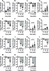
Fig 2. Complementation of the loss of intracellular replication of R. equi 103S ΔvapA with carboxyl-Strep-tag Vaps encoded in the porcine- and the bovine-type virulence plasmids.
J774A.1 macrophage-like cells were infected with virulent R. equi 103S (A), plasmid-cured, R. equi 103SP- (B) and R. equi 103S ΔvapA, an attenuated derivative harbouring an in-frame, unmarked, deletion of vapA (C). The latter was used for complementation analysis using pVapA-ST (R. equi ΔvapA/pVapA-ST) (D) as a positive control and other carboxyl-Strep Tagged vaps from the porcine-type pVAPB1593 (E-J) and bovine-type pVAPN1571 (K-O) virulence plasmids. Infections were set at a MOI of 10 as described in Materials and Methods and the intracellular replication assessed at 24 and 48 hours post infection (HPI) an reported as the Log2 Fold change of CFU per monolayer as compared with time zero. E) R. equi ΔvapA/pVapB-ST; F) R. equi ΔvapA/pVapJ-ST; G) R. equi ΔvapA/pVapK1-ST; H) R. equi ΔvapA/pVapK2-ST; I) R. equi ΔvapA/pVapL-ST; J) R. equi ΔvapA/pVapM-ST; K) R. equi ΔvapA/pVapN-ST; L) R. equi ΔvapA/pVapO-ST; M) R. equi ΔvapA/pVapP-ST; N) R. equi ΔvapA/pVapR-ST; O) R. equi ΔvapA/pVapS-ST. Bars represent the median of at least three independent experiments measured in triplicate. Error bars represent the interquartile range. In some cases, bacteria was cleared during the time course of the experiment. In these cases, data shown only represents values that were possible to count. Statistical analysis was performed using a Kruskal-Wallis test. P-values were adjusted using the Dunn’s correction for multiple comparisons.
Fig 3
Fig 3. Only clade-1 Vap proteins are localised on the cell surface of R. equi.
R. equi 103S ΔvapA transformed with plasmids encoding carboxyl-terminal Strep tagged derivatives of relevant Vaps were stained with the α-Strep tag FITC-mAb and analysed by Flow cytometry. Alive, FITC-stained singlet cells were measured as described in Materials and Methods. The stain index was used to quantify the brightness of stained (positive) cells relative to their unstained (negative) controls. Panels show the log2 results obtained for Vaps encoded by A) the equine type pVAP1037 virulence plasmid, B) the porcine type pVAPB1593 virulence plasmid and C) bovine type pVAPN1571 virulence plasmid (Black symbols). Trypsin digestion before incubation with the α-Strep tag FITC-mAb was employed to confirm the exposure of Strep-tagged proteins to the extracellular environment (Red symbols). Only selected Vaps encoded in the equine type virulence plasmid are shown as example of a protein expressed on the cell surface (VapA-ST) and a protein that is not (VapD-ST), as previously reported. Dot plots of at least three independent experiments are shown. Error bars represent the mean and standard deviation. Statistical analysis was performed in GraphPad using one-way ANOVA. Correction for multiple comparisons was performed using the Šídák test. The p-value of relevant comparisons is depicted above horizontal lines.
Fig 4
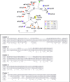
Fig 4. Conservation of the unordered N-terminal Vap domain is clade specific.
A) Unrooted maximum likelihood tree of the Vap protein family encoded by pVAPA (VapA, VapD, VapG, VapH, VapE), pVAPB (VapB, VapK1, VapK2, VapJ, VapL, VapM) and pVAPN (VapN, VapO, VapP, VapR, VapS). The ML tree is based on an alignment of mature Vap proteins, excluding the signal sequence. The number of bootstraps (n = 100) are indicated at the branch points. B) Sequence alignment of the unordered N-terminal sequences of clade-1, clade-2, clade-3, clade-5 and clade-7 proteins. The unordered N-terminal Vap sequences are derived from mature Vap proteins excluding the core Vap proteins as indicated by the structural analysis of VapB, VapD and VapG proteins [–34]. The N-terminal sequences of VapK1 and VapK2 are identical and only included once in the sequence alignment. The VapB N-terminal sequence is excluded from the alignment because it is not a functional VapA homologue, unlike the other clade-1 proteins.

Similar articles

References

    1. <References>. Vázquez-Boland JA, Meijer WG. The pathogenic actinobacterium Rhodococcus equi: what’s in a name? Mol Microbiol. 2019;112(1):1–15. Epub 2019/05/18. doi: 10.1111/mmi.14267 ; PubMed Central PMCID: PMC6852188. - DOI - PMC - PubMed
    1. Vazquez-Boland JA, Giguere S, Hapeshi A, Macarthur I, Anastasi E, Valero-Rello A. Rhodococcus equi: The many facets of a pathogenic actinomycete. Vet Microbiol. 2013;167(1–2):9–33. S0378-1135(13)00330-1 [pii]. - PubMed
    1. Takai S, Watanabe Y, Ikeda T, Ozawa T, Matsukura S, Tamada Y, et al.. Virulence-associated plasmids in Rhodococcus equi. J Clin Microbiol. 1993;31:1726–9. - PMC - PubMed
    1. Ocampo-Sosa AA, Lewis DA, Navas J, Quigley F, Callejo R, Scortti M, et al.. Molecular epidemiology of Rhodococcus equi based on traA, vapA, and vapB virulence plasmid markers. J Infect Dis. 2007;196(5):763–9. - PubMed
    1. Giguère S, Hondalus MK, Yager JA, Darrah P, Mosser DM, Prescott JF. Role of the 85-kilobase plasmid and plasmid-encoded virulence-associated protein A in intracellular survival and virulence of Rhodococcus equi. Infect Immun. 1999;67(7):3548–57. - PMC - PubMed

MeSH terms

LinkOut - more resources