. 2025 Jan 6;16(1):415.
doi: 10.1038/s41467-024-55046-8.

Development of nucleus-targeted histone-tail-based photoaffinity probes to profile the epigenetic interactome in native cells

Affiliations

Development of nucleus-targeted histone-tail-based photoaffinity probes to profile the epigenetic interactome in native cells

Yu Wang et al. Nat Commun. .

Abstract

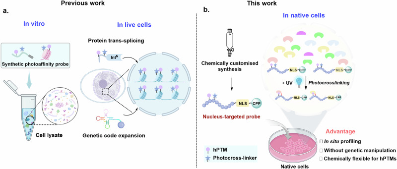
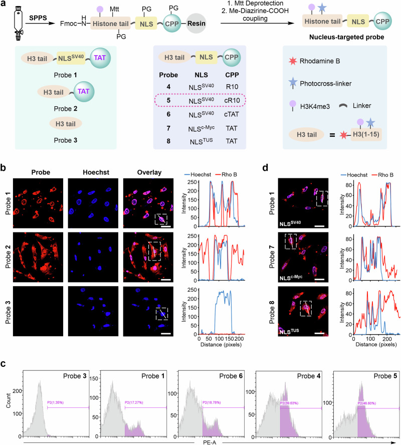
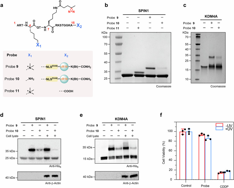
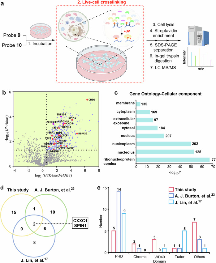
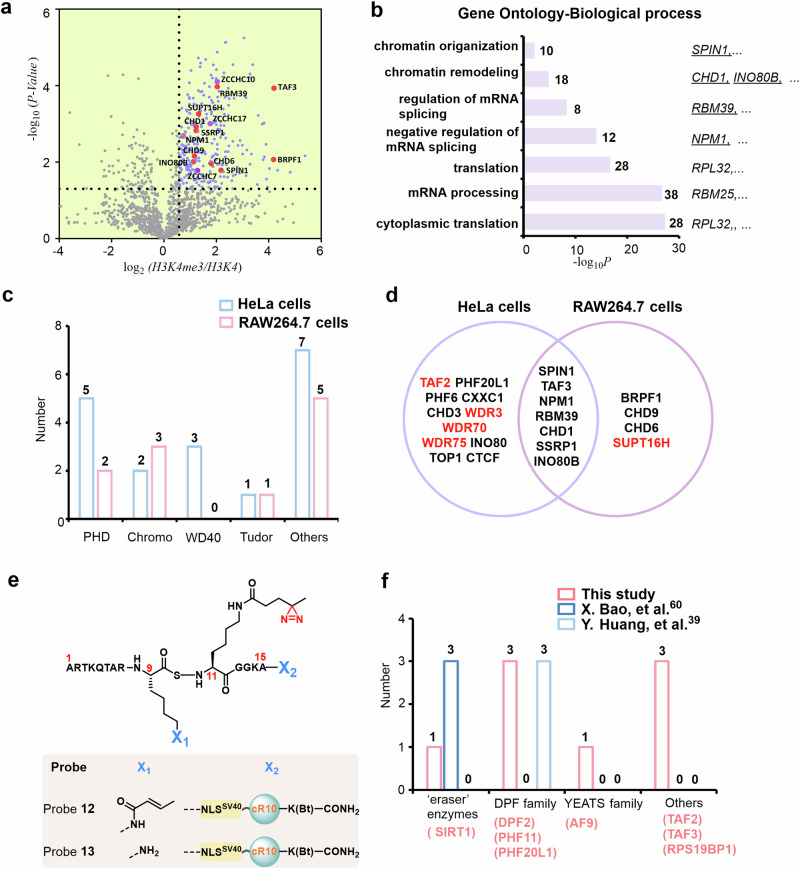
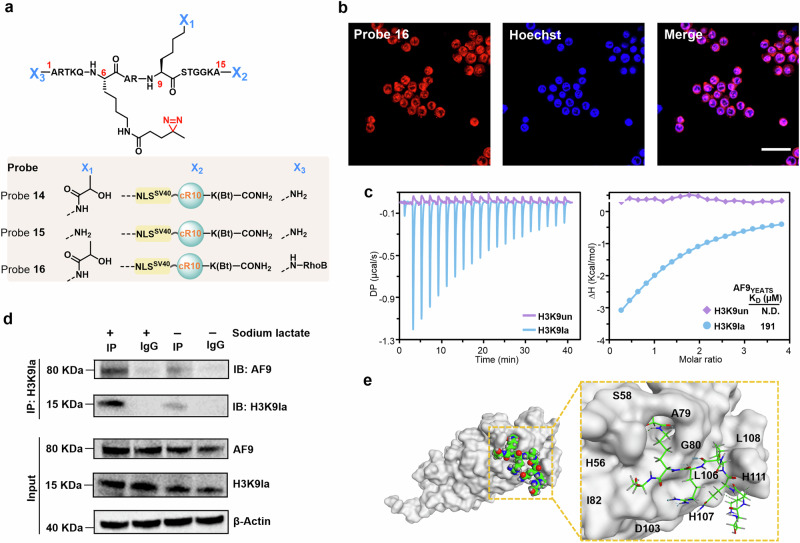
Dissection of the physiological interactomes of histone post-translational modifications (hPTMs) is crucial for understanding epigenetic regulatory pathways. Peptide- or protein-based histone photoaffinity tools expanded the ability to probe the epigenetic interactome, but in situ profiling in native cells remains challenging. Here, we develop a nucleus-targeting histone-tail-based photoaffinity probe capable of profiling the hPTM-mediated interactomes in native cells, by integrating cell-permeable and nuclear localization peptide modules into an hPTM peptide equipped with a photoreactive moiety. These types of probes, such as histone H3 lysine 4 trimethylation and histone H3 Lysine 9 crotonylation probes, enable the probing of epigenetic interactomes both in HeLa cell and hard-to-transfect RAW264.7 cells, resulting in the discovery of distinct interactors in different cell lines. The utility of this probe is further exemplified by characterizing interactome of emerging hPTM, such as AF9 was detected as a binder of histone H3 Lysine 9 lactylation, thus expanding the toolbox for profiling of hPTM-mediated PPIs in live cells.


Conflict of interest statement

Competing interests: The authors declare no competing interests.

Figures

Fig. 1
Fig. 1. Schematic for profiling the hPTM-mediated interactomes.
a Previous strategies that function in an in vitro setting (a test tube) or rely on prior genetic manipulation. b The nucleus-targeted cell-permeable histone-tail-based photoaffinity probes enable it to penetrate into cells and selectively accumulate in the nucleus. The hPTMs-mediated interactomes could be captured in native cells upon UV irradiation. Some elements of Fig. 1a, b were created in BioRender. Chu, G (2024) https://BioRender.com/u07k630.
Fig. 2
Fig. 2. Chemical synthesis and visualization of nucleus-targeted cell-permeable histone-tail-based photoaffinity probes.
a Synthetic scheme and structure of probes 1 to 8. b Confocal microscopy images of HeLa cells treated with probes 1–3 for 1 h at 37 °C. Probes 1–3 were visualized using Rho B fluorescence (red channel), and Hoechst 33258 was utilized for nuclear staining (blue channel). 2D intensity profiles corresponding to the lines displayed on the images (line scans) are depicted to the right of the micrographs. Scale bars: 20 μm. Confocal microscopy images shown in (b) are representative of independent biological replicates (n = 3). c Cellular uptake analysis of probes in HeLa cells by flow cytometry. Cells were treated with probes 1 and 3–6 for 30 min at 37 °C. d Confocal microscopy images and line scans of HeLa cells treated with probes 1, 7, and 8. Scale bars: 20 μm. Confocal microscopy images shown in (d) are representative of independent biological replicates (n = 2). Source data are provided as a Source Data file.
Fig. 3
Fig. 3. Nucleus-targeted histone-tail-based photoaffinity probe to capture readers of H3K4me3 in vitro.
a Chemical structure of probes 9 to 11. b, c SDS-PAGE analysis of the labeling of SPIN1 and KDM4A by probe 9. Probes 10 and 11 were used for comparison for SPIN1 (b), and probe 10 was used for comparison for KDM4A (c). Gel images shown in (b, c) are representative of independent biological replicates (n = 2). d, e Cell lysate profiling using probe 9 with probe 10 as a comparison. Labeled proteins were analyzed by immunoblotting using an anti-His6 antibody. Immunoblotting images shown in (d, e) are representative of independent biological replicates (n = 2). f Viability of HeLa cells treated with 60 μM probe 9. Data were plotted as mean ± SEM of three independent biological replicates.
Fig. 4
Fig. 4. Determining the in situ interactome of H3K4me3 in HeLa cells.
a Schematic for the LFQ proteomics workflow to determine the interactome of H3K4me3 by probe 9. Probe 10 was used for comparison. Some elements of Fig. 4(a) were created in BioRender. Chu, G. (2024) https://BioRender.com/u07k630. b Volcano plots of the quantitative mass spectrometry results. Significantly enriched hits (p < 0.05, >1.5-fold-change) are colored blue. Some established H3K4me3 reader proteins are highlighted and labeled in red. A two-sided test was used. c GO analysis (cellular components) of significantly enriched hits (p < 0.05, >1.5-fold-change) in Fig. 4b. The number of proteins in each GO term is shown. d Venn diagram comparison of identified H3K4me3 reader proteins in the present study and two previous studies, respectively. e Number of identified H3K4me3 reader proteins containing PHD, Chromo, WD40, and Tudor domains in the present study and two previous studies. Source data are provided as a Source Data file.
Fig. 5
Fig. 5. Determining the interactome of H3K4me3 and H3K9cr in RAW264.7 cells.
a Volcano plots of mass spectrometry results. Hits significantly enriched by probe 9 (p < 0.05, >1.5-fold-change) are colored blue. Some established H3K4me3 reader proteins are highlighted and labeled in red. A two-sided test was used. b GO analysis (biological process) of significantly enriched hits (p < 0.05, >1.5-fold-change) in Fig. 5a. The number of proteins in each GO term is shown. c Number of identified H3K4me3 reader proteins containing PHD, chromo, WD40, and Tudor domains by probe 9 in HeLa and RAW264.7 cells. d Overlap of identified H3K4me3 reader proteins using probe 9 in HeLa and RAW264.7 cells. e Chemical structure of probe 12 and probe 13. f Number of identified H3K9cr reader proteins containing eraser enzymes, DPF, YEATS, and other families in the present study and two previous studies. Red-colored words indicate H3K9cr reader proteins enriched in this study. Source data are provided as a Source Data file.
Fig. 6
Fig. 6. Determining the in situ interactome of H3K9la.
a Chemical structure of probes 14, 15, and 16. b Confocal microscopy images of RAW264.7 cells treated with probe 14 for 1 h at 37 °C. Probe 14 was visualized using TER fluorescence (red channel), and Hoechst 33258 was utilized for nuclear staining (blue channel). Scale bars: 20 μm. Confocal microscopy images shown in (b) are representative of independent biological replicates (n = 3). c ITC fitting curves of AF9YEATS with H31-10K9la (blue) peptide or H31-10 (red) peptide. d Co-IP assay was conducted to detect the interaction of H3K9la and AF9 in RAW264.7 cells with or without sodium lactate treatment. Immunoblotting images shown in (d) are representative of independent biological replicates (n = 3). e Structural modeling of AF9YEATS and H31-10K9la peptide. The AF9YEATS structure was displayed on a gray surface, the ligand is shown in sphere and sticks, the carbon atoms are green, the oxygen atoms are red, and the nitrogen atoms are blue. Source data are provided as a Source Data file.

Similar articles

Cited by

References

    1. Kouzarides, T. Chromatin modifications and their function. Cell128, 693–705 (2007). - PubMed
    1. Bannister, A. J. & Kouzarides, T. Regulation of chromatin by histone modifications. Cell Res.21, 381–395 (2011). - PMC - PubMed
    1. Torres, I. O. & Fujimori, D. G. Functional coupling between writers, erasers and readers of histone and DNA methylation. Curr. Opin. Struct. Biol.35, 68–75 (2015). - PMC - PubMed
    1. Lin, J. et al. Menin “reads” H3K79me2 mark in a nucleosomal context. Science379, 717–723 (2023). - PubMed
    1. Boichenko, I. & Fierz, B. Chemical and biophysical methods to explore dynamic mechanisms of chromatin silencing. Curr. Opin. Chem. Biol.51, 1–10 (2019). - PubMed

LinkOut - more resources