

A latent diffusion approach to visual attribution in medical imaging
- PMID: 39762275
- PMCID: PMC11704132
- DOI: 10.1038/s41598-024-81646-x
A latent diffusion approach to visual attribution in medical imaging
Abstract
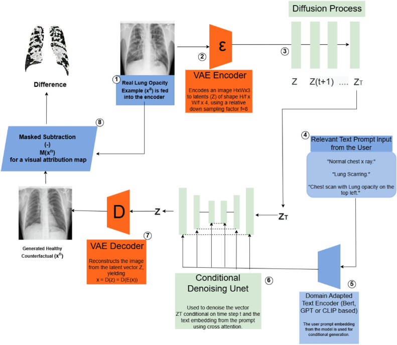
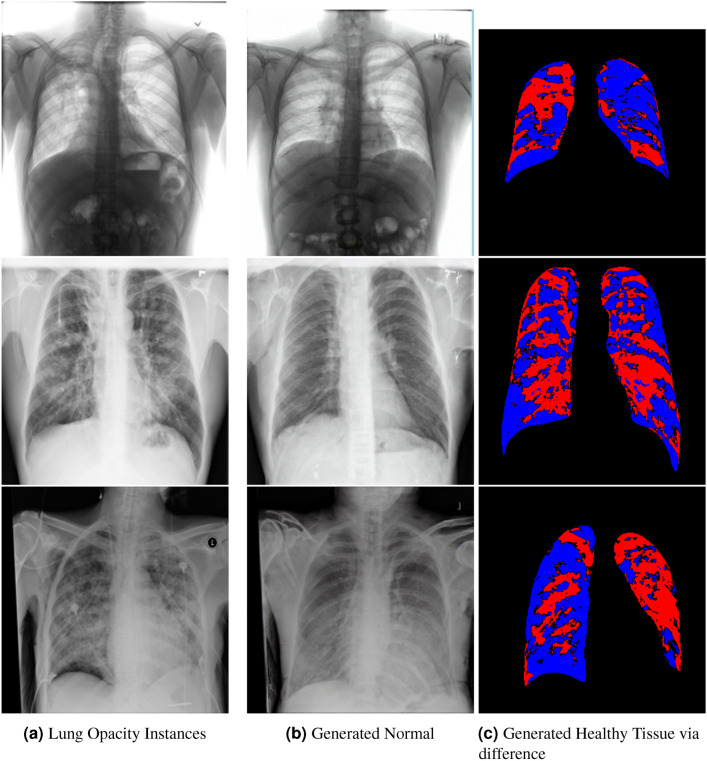
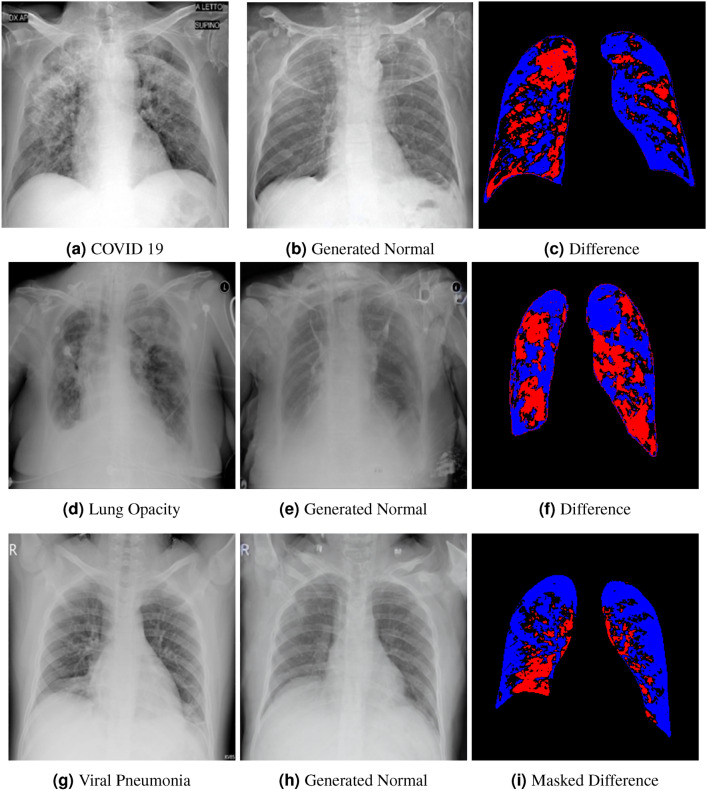
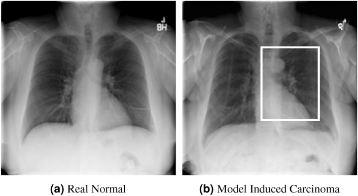
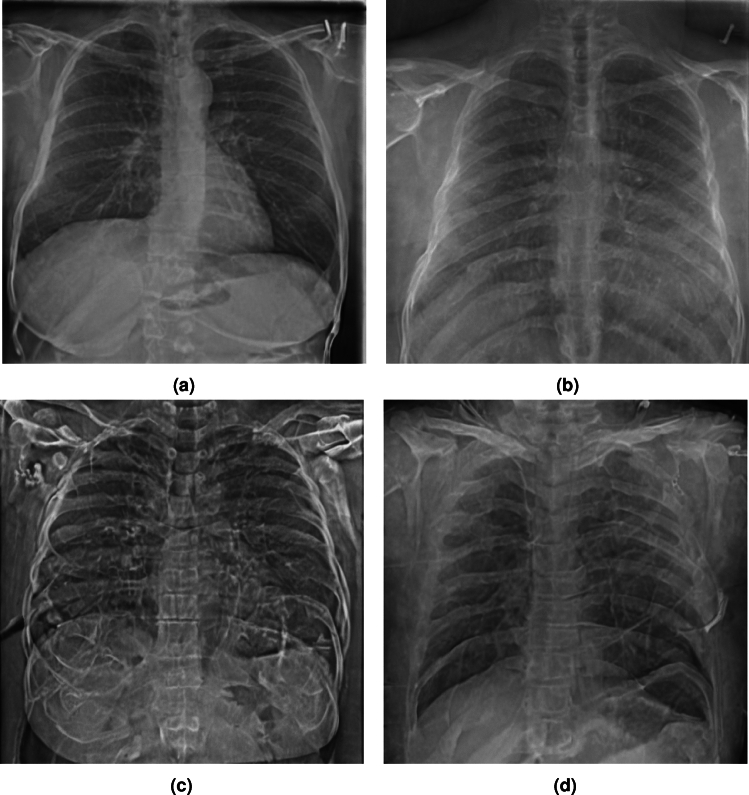
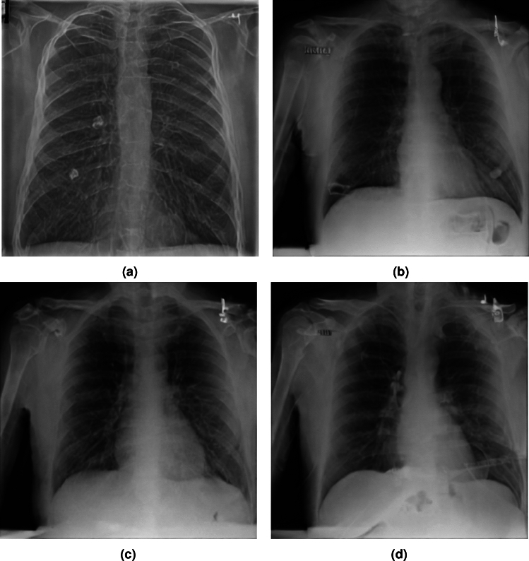
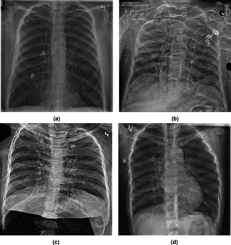
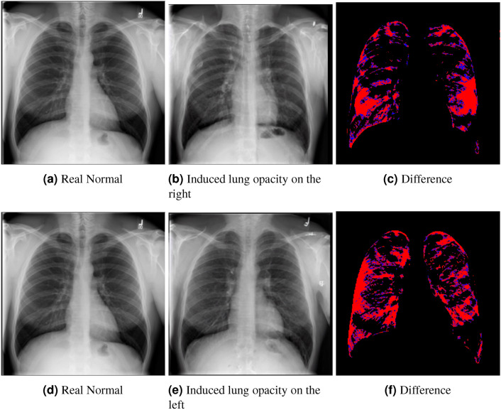
Visual attribution in medical imaging seeks to make evident the diagnostically-relevant components of a medical image, in contrast to the more common detection of diseased tissue deployed in standard machine vision pipelines (which are less straightforwardly interpretable/explainable to clinicians). We here present a novel generative visual attribution technique, one that leverages latent diffusion models in combination with domain-specific large language models, in order to generate normal counterparts of abnormal images. The discrepancy between the two hence gives rise to a mapping indicating the diagnostically-relevant image components. To achieve this, we deploy image priors in conjunction with appropriate conditioning mechanisms in order to control the image generative process, including natural language text prompts acquired from medical science and applied radiology. We perform experiments and quantitatively evaluate our results on the COVID-19 Radiography Database containing labelled chest X-rays with differing pathologies via the Frechet Inception Distance (FID), Structural Similarity (SSIM) and Multi Scale Structural Similarity Metric (MS-SSIM) metrics obtained between real and generated images. The resulting system also exhibits a range of latent capabilities including zero-shot localized disease induction, which are evaluated with real examples from the cheXpert dataset.
Keywords: Diffusion models; Explainable AI; Medical imaging; Visual Attribution.
© 2024. The Author(s).
Conflict of interest statement
Declarations. Competing interests: The authors declare no competing interests.
Figures


References
-
- Baumgartner, C. F., Koch, L. M., Tezcan, K. C., Ang, J. X. & Konukoglu, E. Visual feature attribution using wasserstein gans. In Proceedings of the IEEE conference on computer vision and pattern recognition, 8309–8319 (2018).
-
- Vellido, A., Martín-Guerrero, J. D. & Lisboa, P. J. Making machine learning models interpretable. In ESANN, vol. 12, 163–172 (Citeseer, 2012).
-
- Zhu, W., Lou, Q., Vang, Y. S. & Xie, X. Deep multi-instance networks with sparse label assignment for whole mammogram classification. In International conference on medical image computing and computer-assisted intervention, 603–611 (Springer, 2017).
-
- Ge, Z., Demyanov, S., Chakravorty, R., Bowling, A. & Garnavi, R. Skin disease recognition using deep saliency features and multimodal learning of dermoscopy and clinical images. In Medical Image Computing and Computer Assisted Intervention- MICCAI 2017: 20th International Conference, Quebec City, QC, Canada, September 11-13, 2017, Proceedings, Part III 20, 250–258 (Springer, 2017).
Publication types
MeSH terms
LinkOut - more resources
Full Text Sources
Medical
