. 2025 Jan 6;15(1):978.
doi: 10.1038/s41598-024-84860-9.

Trichostatin A promotes de novo shoot regeneration from Arabidopsis root explants via a cytokinin related pathway

Affiliations

Trichostatin A promotes de novo shoot regeneration from Arabidopsis root explants via a cytokinin related pathway

Su Hyun Park et al. Sci Rep. .

Abstract

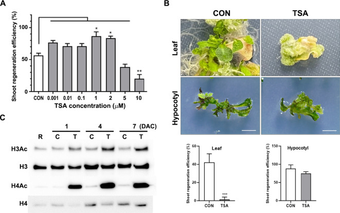
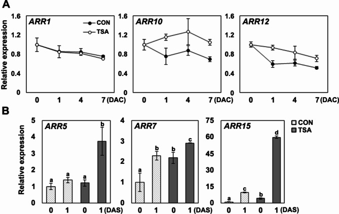
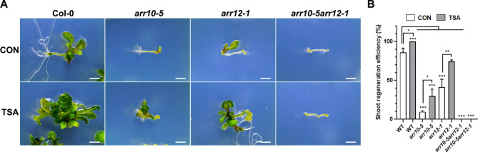
De novo shoot regeneration, characterized by the emergence of adventitious shoots from excised or damaged tissues or organs in vitro, is regulated by the complex interplay between genetic and epigenetic regulatory mechanisms. However, the specific effect of histone deacetylation on shoot regeneration remains poorly understood. This study investigated the effects of trichostatin A (TSA), a histone deacetylase inhibitor, on shoot regeneration in callus derived from root explants. TSA-treated root explants exhibited pronounced callus greening and substantially increasing in multiple shoot formations per callus compared with the control group. Additionally, TSA treatment upregulated shoot apical meristem-specific genes, including WUSCHELL (WUS), RELATED TO AP2.6 L (Rap2.6 L), SHOOT MERISTEMLESS (STM), CUP SHAPED COTYLEDON 2 (CUC2). Notably, TSA treatment enhanced the sensitivity to cytokinins, leading to increase expression of the cytokinin signaling reporter TCS::GFP in the callus. Concomitantly, type-B ARABIDOPSIS RESPONSE REGULATOR (ARR) 10 and 12, which are key regulators of cytokinin signaling, were upregulated in TSA-treated callus, whereas the downstream targets of type-B ARRs, such as ARR5, ARR7, and ARR15, were significantly upregulated during shoot regeneration. Furthermore, mutants deficient in ARR10 and ARR12 showed diminished responsiveness to shoot regenerative capacity, a phenotype that was enhanced by TSA treatment. Our findings underscore the crucial role of histone deacetylation in mediating cytokinin responses and controlling de novo shoot regeneration in plants.

Keywords: Cytokinin response; Histone deacetylation; Shoot regeneration; Trichostatin A.


Conflict of interest statement

Declarations. Competing interests: The authors declare no competing interests. Ethics declarations: Experimental research and field studies on plants, including the collection of plant material, complying with relevant institutional, national, and international guidelines and legislation.

Figures

Fig. 1
Fig. 1
TSA promotes shoot regenerative capacity. (A) Efficiency of shoot regeneration in callus treated with various TSA concentrations. Root explants from 7-day-old seedlings were cultured in CIM with indicated concentration of TSA, then transferred to SIM without TSA and incubated for 18 days under continuous light. Data are expressed as means ± standard deviation of three biological replicates. Significant differences were determined using one-way ANOVA followed by Tukey’s post hoc test, with distinct letters indicating p < 0.05. (B) Upper panels: shoot regeneration of leaf and hypocotyl explants incubated in CIM with or without 2 µM TSA. Hypocotyl explants 7-day-old seedling and leaf explants from 14-day-old seedlings were cultured in CIM, followed by a 4-week incubation in SIM under continuous light. Scale bar = 2 mm. Lower panels: shoot regeneration efficiency of leaf and hypocotyl explants treated with or without 2 µM TSA. Data are expressed as mean ± standard deviation. Statistical significance was assessed using Student’s t-test, with ***p < 0.001. (C) Effect of TSA on histone H3 and H4 acetylation levels during callus induction. Histone proteins were extracted from 7-day-old seedling root explants with callus incubated in CIM with or without TSA after 1, 4, and 7 days. R: Root, C: control callus, T: TSA-treated callus, DAC: days after CIM incubation. Immunoblot images are cropped from supplementary information (Fig. S1).
Fig. 2
Fig. 2
Effect of TSA onde novoshoot regeneration from callus derived from root explants. (A) Shoot regeneration of TSA-treated and control calli. Root explants from 7-day-old seedlings were cultured in CIM with or without 2 µM TSA for 7 days in the dark, then transferred to shoot induction medium (SIM) without TSA for 21 days under continuous light. DAS indicates days after SIM incubation. Scale bars = 2 mm. (B) Efficiency of shoot regeneration for TSA-treated and control calli, measured on designated days. (C) Number of regenerated shoots per explant for TSA-treated and control calli. The number of shoots was measured three weeks after transfer to SIM. Data represent means ± standard deviation of three biological replicates. Statistical significance was assessed using Student’s t-test, with significance levels indicated as ***p < 0.001.
Fig. 3
Fig. 3
Effect of TSA on the expression of shoot meristem-specific genesAtWUS, AtSTM, AtRap2.6 L, andAtCUC2during shoot regeneration. Calli were induced from root explants in CIM with or without 2 µM TSA for 7 days, then transferred to SIM without TSA. Gene expression was normalized to eIF4A. Data represent the means of three biological replicates, with error bars showing standard deviation. Significant differences between TSA concentrations were determined using one-way ANOVA with Tukey’s post hoc test, indicated by distinct letters (p < 0.05).
Fig. 4
Fig. 4
TSA confers cytokinin hypersensitivity during shoot regeneration. (A) Shoot regeneration efficiency of callus treated with TSA at various 2-iP concentrations. Root explants from 7-day-old seedlings were cultured in CIM with or without 2 µM TSA, then transferred to SIM containing different concentrations of 2-iP under continuous light. Photos were taken two weeks after the transfer. Scale bar = 2 mm. Statistical significance between control and TSA-treated groups at each concentration was determined using Student’s t-test, and the results are presented as mean ± standard deviation (***p < 0.001). (B) Effect of TSA on cytokinin signaling using TCS::GFP reporter. Callus treated with or without TSA was transferred to SIM and imaged by confocal microscopy. Callus was stained with 10 µM propidium iodide to visualize cell outlines; scale bar = 50 μm.
Fig. 5
Fig. 5
Effect of TSA on expression of cytokinin signaling during callus induction. Root explants taken from seedling of 7 days old were cultured in CIM treated with or without 2 µM TSA. After 7 days incubation, the calli were harvested to extract total RNA for quantitative RT-PCR analysis. Gene expression was adjusted to be comparable to that of the eIF4A gene. Each value indicates means of three biological replicates and error bars show standard deviation. Statistical significance was determined using student t-test (*p < 0.05, **p < 0.01 and ***p < 0.001).
Fig. 6
Fig. 6
Effect of TSA on the expression of cytokinin signaling regulators during shoot regeneration. (A) Expression profiles of ARR1, ARR10, and ARR12 during callus induction with TSA treatment. Root explants from 7-day-old seedlings were cultured in CIM with or without TSA, and callus was harvested at indicated days for RNA extraction. (B) Relative expression levels of ARR5, ARR7, and ARR15 during shoot regeneration. Callus was induced from root explants in CIM with or without TSA for 7 days, then transferred to SIM. RNA was extracted from callus incubated for 7 days in CIM (0 DAS) and 7 days in CIM plus 1 day in SIM (1 DAS). Transcript levels were normalized to eIF4A. Data represent means of three biological replicates, with error bars showing standard deviation. Significant differences between treatments were determined using one-way ANOVA with Tukey’s post hoc test, indicated by distinct letters (p < 0.05).
Fig. 7
Fig. 7
Effect of TSA on shoot regeneration ofarr10-5, arr12-1, andarr10-5arr12-1mutants. (A) Root explants from 7-day-old seedlings were incubated on CIM with 2 µM TSA for 7 days then transferred to SIM without TSA. Photos were taken 3 weeks after transfer. The scale bar = 2 mm. (B) Shoot regeneration efficiency of arr10-5, arr12-1, and arr10-5arr12-1 mutants treated with or without 2 µM TSA. Data are presented as mean ± standard deviation (n = 3 samples) and are representative of three independent experiments. Statistical significance was assessed using Student’s t-test, with *p < 0.05, **p < 0.01, and ***p < 0.001.

References

    1. Christianson, M. L. & Warnick, D. A. Competence and determination in the process of in vitro shoot organogenesis. Dev. Biol.95, 288–293. 10.1016/0012-1606(83)90029-5 (1983). - PubMed
    1. Mayer, K. et al. Role of WUSCHEL in regulating stem cell fate in the Arabidopsis shoot meristem. Cell95, 805–815. 10.1016/S0092-8674(00)81703-1 (1999). - PubMed
    1. Barton, M. & Poethig, R. S. Formation of the shoot apical meristem in Arabidopsis thaliana—An analysis of development in wild type and in the SHOOT MERISTEMLESS mutant. Development119 (1993).
    1. Chatfield, S. P. et al. Incipient stem cell niche conversion in tissue culture: Using a systems approach to probe early events in WUSCHEL-dependent conversion of lateral root primordia into shoot meristems. Plant. J.73, 798–813. 10.1111/tpj.12085 (2013). - PubMed
    1. Gordon, S. P. et al. Pattern formation during de novo assembly of the Arabidopsis shoot meristem. Development134, 3539–3548. 10.1242/dev.010298 (2007). - PubMed

Publication types

MeSH terms

Grants and funding

LinkOut - more resources