

A case of large uterine cystic adenomyosis outside the uterus after laparoscopic myomectomy: a case report and literature review
- PMID: 39762785
- PMCID: PMC11705667
- DOI: 10.1186/s12905-024-03543-9
A case of large uterine cystic adenomyosis outside the uterus after laparoscopic myomectomy: a case report and literature review
Abstract
Background: Uterine cystic adenomyosis is a rare form of focal adenomyosis that is primarily located within the myometrium. In this case report, we present a unique case of adult uterine cystic adenomyosis found outside the uterus following laparoscopic myomectomy.
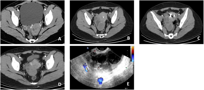
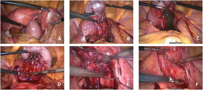
Case presentation: The patient was a 36-year-old Chinese woman who had previously undergone laparoscopic surgery at our hospital to remove a 4 cm diameter diameter uterine fibroid six years prior. She returned to our hospital due to excessive menstruation and intermittent right lower abdominal pain for one year. Pelvic computed tomography revealed an endometriotic cyst on the right posterior side of the uterus seemingly connected to the uterine cavity. During surgery, we successfully removed a large well-defined cyst filled with typical chocolate-like fluid located outside the uterus with its root deeply embedded in the myometrium. Following surgery, we administered gonadotropin-releasing hormone analogs for six cycles without any observed recurrence.
Conclusion: This study describes an unusual occurrence of large adult uterine cystic adenomyosis located outside the uterus after laparoscopic myomectomy, potentially supporting the theory that endometrial injury invagination may be responsible for this condition.
Keywords: Laparoscopic myomectomy; Uterine cystic adenomyosis; Uterine fibroids.
© 2024. The Author(s).
Conflict of interest statement
Declarations. Ethics approval and consent to participate: This paper was approved by the Ethics Committee of the institutional review board (IRB) of Shengjing Hospital of China Medical University. The patient described in this case report provided informed consent. Consent for publication: Written informed consent was obtained from the patient for the publication of this case report and any accompanying images. Competing interests: The authors declare no competing interests.
Figures
References
-
- Cullen TS. Adenomyoma of the uterus. Philadelphia: Saunders; 1908. pp. 128–49.
Publication types
MeSH terms
LinkOut - more resources
Full Text Sources
Medical
