. 2025 Jan 7;20(1):1.
doi: 10.1186/s13000-024-01576-0.

Gene expression and immunohistochemistry analysis of ADAMTS-1 and its substrates in odontogenic keratocyst

Affiliations

Gene expression and immunohistochemistry analysis of ADAMTS-1 and its substrates in odontogenic keratocyst

Osvaldo Rodrigues de Souza Neto et al. Diagn Pathol. .

Abstract

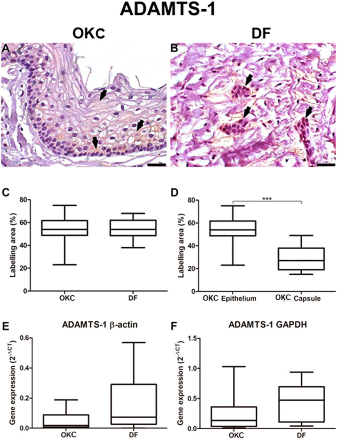
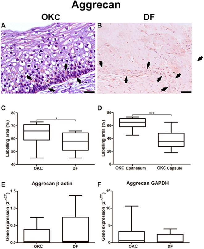
Background: Considering the significant participation of the microenvironment in the local aggressiveness of odontogenic keratocysts, this study aims to evaluate the expression of ADAMTS-1 and its substrates, versican, aggrecan and brevican in this locally invasive odontogenic cyst.

Methods: Immunohistochemistry and polymerase chain reaction (PCR) were conducted on 30 cases of odontogenic keratocysts (OKCs) and 20 dental follicles (DFs).

Results: The immunohistochemical expression of these proteins was predominantly cytoplasmic and granular across all samples. In epithelial tissue, the immunoexpression of aggrecan and versican was higher in OKC (p < 0.05) compared to DF. Comparing the expression of proteins between the OKC epithelium and the cystic capsule, it was observed that all molecules were more expressed in the epithelium (p < 0.001). RT-PCR confirmed the expression of ADAMTS-1 and proteoglycans in all samples.

Conclusion: ADAMTS-1, aggrecan, brevican, and versican were expressed in all samples with a granular and cytoplasmic pattern. RT-PCR confirmed their presence in both OKC and DF, but only aggrecan and versican exhibited significantly higher levels in OKC (p < 0.05). Protein expression was notably greater in the epithelial component of OKC. These findings underscore the potential role of these proteins in the biological behavior of OKC.

Keywords: ADAMTS-1; Immunohistochemistry; Odontogenic keratocyst; Proteoglycans.


Conflict of interest statement

Declarations. Ethics approval and consent to participate: this study was approved by the Ethics Committee of the Institute of Oncology Research Center of the Federal University of Pará (n°2.371.646). Consent was obtained from patients for use of their samples. Consent for publication: not applicable. Competing interests: The authors declare no competing interests. Declaration of generative AI and AI-assisted technologies in the writing process: During the preparation of this work the authors used ChatGPT (version GPT-4) only to improve readability and language. After using this tool, the authors reviewed and edited the content as needed and take full responsibility for the content of the publication.

Figures

Fig. 1
Fig. 1
ADAMTS-1 immunostaining in odontogenic keratocyst (OKC) and dental follicle (DF). ADAMTS-1 staining was diffuse in the epithelial layers of OKC, with a predominantly cytoplasmic localization (A, arrows). DF also expressed ADAMTS-1, with predominantly cytoplasmic staining (B, arrows). There was no significant difference in the area of ADAMTS-1 staining between OKC and DF (C). However, ADAMTS-1 immunostaining in OKC was more pronounced in epithelial cells than in the capsule (D). ADAMTS-1 gene expression was reduced in OKC compared with DF, although without a statistically significant difference. RT-PCR analysis was performed using the Pfaffl method to calculate relative mRNA levels normalized by β-actin (E) or GAPDH (F). Significance: ∗∗∗p < 0.001. Scale bar: 20 μm
Fig. 2
Fig. 2
Aggrecan immunostaining in odontogenic keratocyst (OKC) and dental follicle (DF). Aggrecan staining was predominantly cytoplasmic in the basal and parabasal layers of OKC epithelial cells (A, arrows). DF exhibited weak aggrecan labeling, primarily cytoplasmic (B, arrows). The labeling area of aggrecan showed higher expression in the epithelial cells of OKC compared to DF (C). Moreover, aggrecan immunostaining in OKC was more pronounced in the epithelial cells than in the capsule (D). There was no statistically significant difference in aggrecan gene expression between OKC and DF, as determined by RT-PCR analysis using the Pfaffl method to calculate relative mRNA levels normalized by β-actin (E) or GAPDH (F). Significance: *p < 0.05, ∗∗∗p < 0.001. Scale bar: 20 μm
Fig. 3
Fig. 3
Brevican immunostaining in odontogenic keratocyst (OKC) and dental follicle (DF). Brevican staining was diffuse in the epithelial layers of OKC, with predominantly cytoplasmic localization (A, arrows). DF also expressed brevican, with predominantly cytoplasmic staining (B, arrows). There was no significant difference in brevican labeling area between OKC and DF (C). However, brevican immunostaining in OKC was more pronounced in the epithelial cells than in the capsule (D). There was no statistically significant difference in brevican gene expression between OKC and DF, as determined by RT-PCR analysis using the Pfaffl method to calculate relative mRNA levels normalized by β-actin (E) or GAPDH (F). Significance: ∗∗∗p < 0.001. Scale bars: 20 μm and 50 μm
Fig. 4
Fig. 4
Versican immunostaining in odontogenic keratocyst (OKC) and dental follicle (DF). Versican staining was predominantly cytoplasmic in the basal and parabasal layers of OKC epithelial cells (A, arrows). DF showed weak versican labeling, with predominantly cytoplasmic localization (B, arrows). The versican labeling area indicated higher expression in the epithelial cells of OKC compared to DF (C). Moreover, versican immunostaining in OKC was more prominent in the epithelial cells than in the capsule (D). There was no statistically significant difference in versican gene expression between OKC and DF, as determined by RT-PCR analysis using the Pfaffl method to calculate relative mRNA levels normalized by β-actin (E) or GAPDH (F). Significance: *p < 0.05, ∗∗∗p < 0.001. Scale bar: 20 μm
Fig. 5
Fig. 5
Signaling pathway between ADAMTS-1, Versican, Aggrecan in the context of keratocysts

Similar articles

References

    1. Pogrel MA. (2013). The keratocystic odontogenic tumor. Oral and maxillofacial surgery clinics of North America, 25(1), 21–30, v. 10.1016/j.coms.2012.11.003 - PubMed
    1. Kaczmarzyk T, Stypułkowska J, Tomaszewska R. Update of the WHO classification of odontogenic and maxillofacial bone tumors. J Stomatology. 2017;70(5):484–506.
    1. WHO Classification of Tumors Editorial Board. Head and Neck Tumours. 5th ed. Lyon: IARC; 2024.
    1. Stoelinga PJW. Keratocystic odontogenic tumour (KCOT) has again been renamed odontogenic keratocyst (OKC). Int J Oral Maxillofac Surg. 2019;48(3):415–6. - PubMed
    1. Mendes RA, Carvalho JF, van der Waal I. Characterization and management of the keratocystic odontogenic tumor in relation to its histopathological and biological features. Oral Oncol. 2010;46(4):219–25. 10.1016/j.oraloncology.2010.01.012. - PubMed

LinkOut - more resources