. 2025 Jan 6;17(1):7.
doi: 10.1186/s13195-024-01648-9.

Neuroprotection by chronic administration of Fluoroethylnormemantine (FENM) in mouse models of Alzheimer's disease

Affiliations

Neuroprotection by chronic administration of Fluoroethylnormemantine (FENM) in mouse models of Alzheimer's disease

Allison Carles et al. Alzheimers Res Ther. .

Abstract

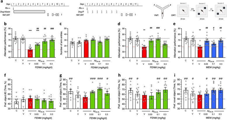
Background: Fluoroethylnormemantine (FENM), a new Memantine (MEM) derivative, prevented amyloid-β[25-35] peptide (Aβ25-35)-induced neurotoxicity in mice, a pharmacological model of Alzheimer's disease (AD) with high predictive value for drug discovery. Here, as drug infusion is likely to better reflect drug bioavailability due to the interspecies pharmacokinetics variation, we analyzed the efficacy of FENM after chronic subcutaneous (SC) infusion, in comparison with IP injections in two AD mouse models, Aβ25-35-injected mice and the transgenic APPswe/PSEN1∂E9 (APP/PS1) line.

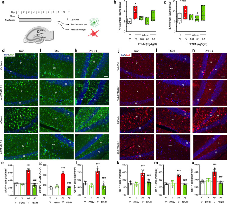
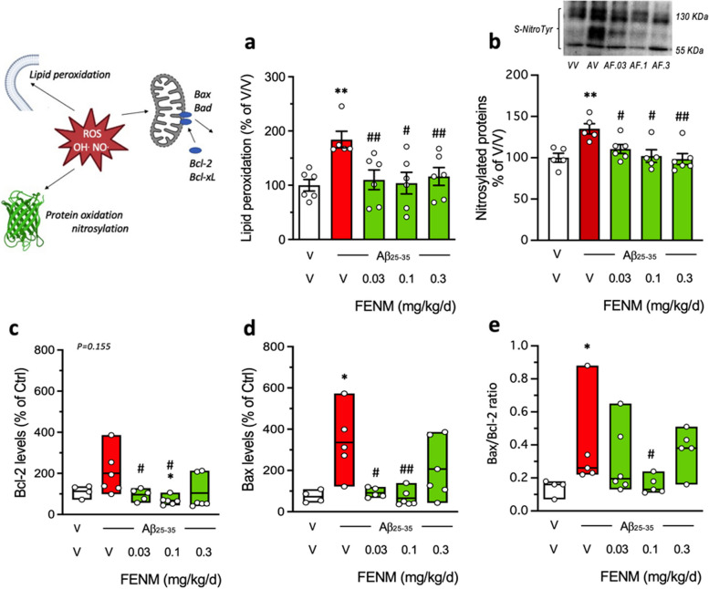
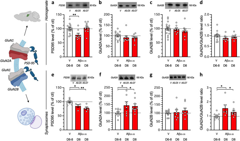
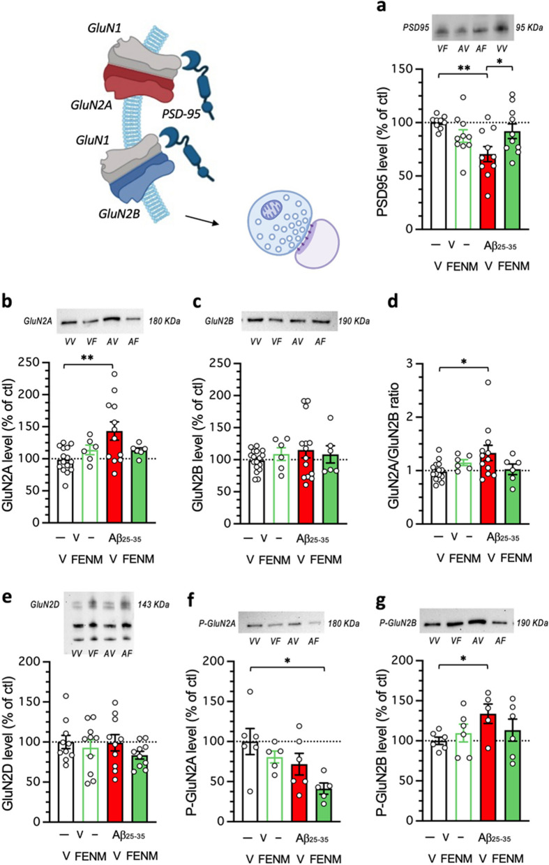
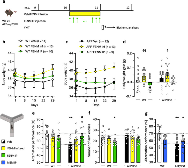
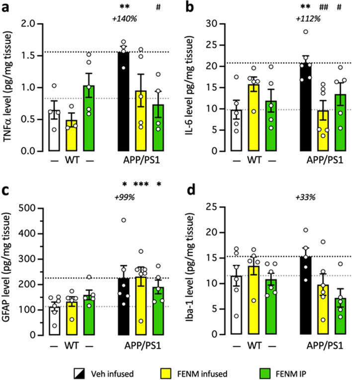
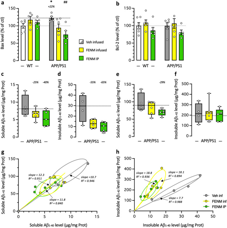
Methods: In Aβ25-35-treated mice, FENM was infused at 0.03-0.3 mg/kg/day during one week after Aβ25-35 injection. For comparison, FENM and MEM were administered IP daily at 0.03-0.3 mg/kg. In 10-month-old APP/PS1 mice, FENM was administered during four weeks by daily IP injections at 0.3 mg/kg or chronic SC infusion at 0.1 mg/kg/day. Memory deficits, spatial working memory and recognition memory, were analysed. Markers of neuroinflammation, apoptosis, oxidative stress, and amyloid burden in APP/PS1 mice, were quantified. Markers of synaptic plasticity such as PSD-95 and GluN2A/B/D subunits expression in hippocampus homogenates or synaptosomes were quantified in Aβ25-35-treated mice and synaptic long-term potentiation (LTP) in hippocampal slices was analysed in APP/PS1 mice.

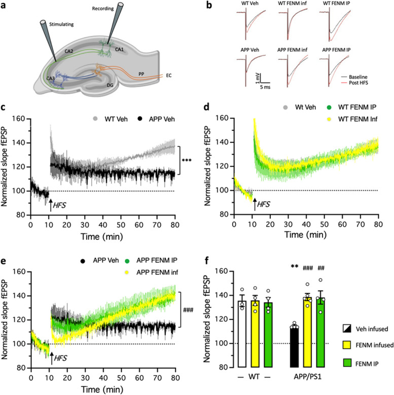
Results: Deficits in spontaneous alternation and object recognition in Aβ25-35 mice were prevented by infused FENM at all doses tested. Similar effects were observed with the daily FENM or MEM treatments. Animals infused with 0.1 mg/kg/day FENM showed prevention of Aβ25-35-induced neuroinflammation, oxidative stress and apoptosis. FENM infusion restored Aβ25-35-induced alterations in synaptosomal PSD-95, GluN2A and P-GluN2B levels. GluN2D levels were unchanged whatever the treatment. In APP/PS1 mice, FENM infused or administered IP alleviated spontaneous alternation deficits, neuroinflammation, increases in Aβ1-40/Aβ1-42 and hippocampal LTP alteration.

Conclusion: These data confirmed the neuroprotective potential of FENM in the pharmacological Aβ25-35 and transgenic APP/PS1 mouse models of AD, with a superiority to MEM, and showed that the drug can be efficiently infused chronically.

Keywords: Alzheimer's disease; Drug infusion; Fluoroethylnormemantine (FENM); NMDA receptor expression; Neuroprotection.


Conflict of interest statement

Declarations. Ethics approval and consent to participate: Animal procedures were conducted in adherence with the European Union Directive 2010/63 and the ARRIVE guidelines and authorized by the National Ethic Committee (Paris, France): authorization APAFIS #30410–2021031516372048. Competing interests: A.F. is employee of ReST Therapeutics. G.R. is co-inventor and owner of the patent FR2005138 (2022) and founder of ReST Therapeutics. T.M. is co-inventor of the patent FR2005138. Other authors declare that they have no conflict of interest to disclose.

Figures

Fig. 1
Fig. 1
Protective effect of SC-infused Fluoroethylnormemantine (FENM) or IP-administered FENM or Memantine (MEM) on Aβ25-35-induced memory impairments in mice. a Experimental protocols for the SC drug infusion or drug IP administration. Abbreviations: YMT, Y-maze test; ORT, object recognition test; †, sacrifice before immunofluorescence or biochemical analyses. Spontaneous alternation in the Y-maze consists in recording the successive arm exploration in the maze over 8 min. The object recognition test consists in three 10-min duration sessions (i) without objects, (ii) with two identical objects, and (iii) with one familiar and one novel object. The object exploration preference was determined on the duration of contacts with each object Animals received Aβ25-35 (9 nmol ICV) on day 1 and then FENM (0.03-0.3 mg/kg/day in the Alzet pump) or FENM or MEM (0.03-0.3 mg/kg IP o.d.) between day 1 to 7. b, d, e spontaneous alternation deficits and (c) number of arm entries. f session 2 of the object recognition test with two identical objects and (g, h, i) session 3 with a novel and a familiar object. Exploration preferences are calculated with the duration of contacts. Abbreviations: C, control (untreated) animals; V, vehicle solution (distilled water for ICV and physiological saline for IP injections of SC infusion. Graphs show individual data and mean ± SEM of N = 7-12 animals per group. Dotted lines show the (V + V)- and (V + Aβ25-35)-treated group levels in (b-e) and the 50% preference level in (f-i). Abbreviations: C, control, non-injected animals; V, vehicle solution. * p < 0.05, ** p < 0.01, *** p < 0.001 vs. (V + V)-treated group; ## p < 0.01, ### p < 0.001 vs. (V + Aβ25-35)-treated group; Dunnett’s test. @ p < 0.05, @@ p < 0.01, @@@ p < 0.001 vs. 50% level, one-sample t-test
Fig. 2
Fig. 2
Protective effects of SC-infused FENM, 0.1 mg/kg/day, on Aβ25-35-induced neuroinflammation in mice. a Experimental protocol and hippocampal areas of interest. b TNFα and (c) IL-6 levels were analyzed in homogenates by ELISA. d-i Astroglial reaction was analyzed in the hippocampus of Aβ25-35-treated mice using GFAP immunolabeling. j-o Microglial reaction in the hippocampus of Aβ25-35-treated mice using Iba-1 immunolabeling. d, e, j, k stratum radiatum (Rad), f, g, l, m stratum lacunosum-moleculare (Mol) and (h, i, n, o) polymorphic layer of the dentate gyrus (PoDG) with (d, f, h, j, l, n) typical immunofluorescence micrographs (blue: DAPI, green: GFAP, red: Iba-1) and (e, g, i, k, m, o) quantifications. Coronal 25 µm thick sections were stained with anti-GFAP or anti-Iba-1 antibody and the three areas of the hippocampus analyzed. Abbreviations: sp, stratum pyramidale; sr, stratum radiatum; sg, stratum granulare; sm, stratum lacunosum-moleculare; Hi, hilus. Scale bar in (h, n) = 50 µm applying to all pictures. The number of slice analyzed per mouse was n = 5–6 with N = 5 mice per group. *** p < 0.001 vs. the (V + V)-treated group; ### p < 0.001 vs. the (Aβ25-35 + V)-treated group; Dunnett's test
Fig. 3
Fig. 3
Protective effects of SC infusion of FENM, on Aβ25-35-inducedoxidative stress and apoptotic markers. a lipid peroxidation, b nitrated proteins, c Bcl-2 level, d Bax level, and (e) Bax/Bcl-2 ratio, measured by colorimetric assay, western blotting or ELISA in mouse hippocampus extracts. Animals were sacrificed 11 days after Alzet pump implantation. In (b), data show mean ± SEM and a typical blot is shown above the graph. In (c-e), data show median and min/max with individual measures. The number of animals per group was N = 4–6. * p < 0.05, ** p < 0.01 vs. (V + V)-treated group; # p < 0.05, ## p < 0.01 vs. (V + Aβ25-35)-treated group; Dunn's test
Fig. 4
Fig. 4
Impact of Aβ25-35 toxicity on PSD-95 and NMDAR GluN2A/B subunits in the mouse hippocampus. Aβ25-35 was administered ICV on day 1 and after 5 or 7 days, the hippocampus was dissected out and protein expression analyzed in homogenates (a, b, c, d) or synaptosomes preparations (e, f, g, h) by western blot: PSD-95 levels (a, e), GluN2A expression (b, f), GluN2B expression (c, g) and GluN2A/GluN2B ratios (d, h). Note that protein levels were not altered on days 1 or 3 after Aβ25-35 peptide ICV injection (data not shown). Vehicle solution (V)-treated animals were sacrificed at day 6 or 8 and data were pooled. Typical blots are shown above each graph. The whole immunostained and and stain-free blots are shown in Supplementary Figs. 1 and 2. The number of animals per group was N = 8–16 per group in (a-d) and N = 4–11 in (e–h). * p < 0.05, ** p < 0.01; Dunnett's test
Fig. 5
Fig. 5
Protective effects of a SC infusion of FENM (0.1 mg/kg/day), on Aβ25-35-induced alteration of PSD-95 and NMDAR subunits expressions in synaptosome preparations of mouse hippocampus: a PSD-95, b GluN2A, c GluN2B, d GluN2A/GluN2B ratio, e GluN2D, f P(Tyr1325)-GluN2A, g P(Ser1303)-GluN2B. Animals were sacrificed 7 days after the peptide injection. Typical blots are shown above each graph. The whole immunostained and and stain-free blots are shown in Supplementary Figs. 3 and 4. The number of animals per group was N = 5–17 per group. * p < 0.05, ** p < 0.01; Dunnett's test. Illustrations were done with www.biorender.com
Fig. 6
Fig. 6
a Experimental protocol. Wildtype (WT) or APPswe/PS1∂E9 (APP/PS1) mice were infused SC with vehicle solution (Veh) or FENM (0.1 mg/kg/day) or repeatedly injected IP with FENM (0.3 mg/kg) or MEM (0.3 mg/kg) between 10 to 11 month-of-age (m.o.). Surgery was performed on day 1, infusion lasted 28 days, and a Y-maze test (YMT) was performed on day 30. On day 31, animals were sacrificed (†) before biochemical analyses. b,c body weight measured weekly for WT (b) and APP/PS1(c) treated groups. d Daily weight gain between day 1 to day 29. § p < 0.05, §§ p < 0.01 vs. 0 (one-column t-test). e Spontaneous alternation performances and (f) total numbers of arm entries for FENM-treated groups and (g) spontaneous alternation performances for MEM-treated groups, in the Y-maze test. * p < 0.05, ** p < 0.01 vs. Veh-treated WT group; # p < 0.05 vs. Veh-treated APP/PS1 group; Dunnett's test
Fig. 7
Fig. 7
Protective effects of FENM on neuroinflammation in the hippocampus of APP/PS1 mice. Vehicle solution (Veh) or FENM (0.1 mg/kg/day) was infused for 4 weeks SC or FENM (0.3 mg/kg) was injected IP o.d. for 4 weeks. animals were sacrificed 48 h after the last administration day. Levels of the cytokines TNFα (a) or IL-6 (b) and the glial markers GFAP (c) or Iba-1 (d) were analyzed in homogenates by ELISA. Data were expressed as percentage of the control, Veh-treated WT group. The number of animal per group was n = 5–6. * p < 0.05, ** p < 0.01, *** p < 0.001 vs. the Veh-treated WT group; # p < 0.05, ## p < 0.01 vs. the Veh-treated APP/PS1 group; Dunnett's test
Fig. 8
Fig. 8
Protective effects of FENM on neuroinflammation in the cortex of APP/PS1 mice. Vehicle solution (Veh) or FENM (0.1 mg/kg/day) was infused for 4 weeks SC or FENM (0.3 mg/kg) was injected IP o.d. for 4 weeks. animals were sacrificed 48 h after the last administration day. Levels of the pro-apoptotic marker Bax (a), the anti-apoptotic marker Bcl-2 (b), and the soluble (c, e) and insoluble (d, f) contents in Aβ1-40 (c, d) and Aβ1-42 (e, f) were analyzed in homogenates by ELISA. Correlations between Aβ1-40 and Aβ1-42 levels are shown in (g) for soluble extracts and in (h) for insoluble extracts. Data in (a, b) were expressed as percentage of the control, Veh-treated WT group. Data in (c-f) were expressed as pg/mg of tissue and represented as box-and-whiskers showing the median and range. The number of animal per group was n = 4–6. * p < 0.05 vs. the Veh-treated WT group; ## p < 0.01 vs. the Veh-treated APP/PS1 group; Dunnett's test
Fig. 9
Fig. 9
Recovery of LTP in the CA1 hippocampal area in APP/PS1 mice by FENM treatments. a Schematic illustration of field excitatory postsynaptic potential (fEPSP) recordings in hippocampal slices. Abbreviations: CA1 ~ 3, Cornus Ammona 1 ~ 3 layers; DG, dentate gyrus; EC, entorhinal cortex; PP, perforant path. b Typiical examples of the fEPSPs recorded in the CA1 area in acute mouse hippocampal slices before (black) and 60 min after (red) high frequency stimulations (HFS). c-e The mean amplitude of fEPSPs, expressed as a percentage of the baseline level, is plotted for: c the Veh-treated WT and APP/PS1 groups; d the Veh-treated, FENM-infused and FENM repeatedly IP injected WT groups; and (e) the Veh-treated, FENM-infused and FENM repeatedly IP injected APP/PS1 groups. It shows the 10 min of baseline recording and 70 min of post‑HFS (arrow) recording. Responses were evoked and collected every 15 s. f Average amplitude of fEPSPs during the the last 10 min post‑HFS recorded in WT and APP/PS1 mice. Data are presented as the mean ± SEM, n = 4–5 animals per group, ** p < 0.01, *** p < 0.001 vs. Veh-treated WT group; ## p < 0.01, ### p < 0.001 vs. Veh-treated APP/PS1 group; repeated-value one-way ANOVA in (c, e), Mann–Whitney test in (f)

References

    1. Brookmeyer R, Gray S, Kawas C. Projections of Alzheimer’s disease in the United States and the public health impact of delaying disease onset. Am J Public Health. 1998;88:1337–42. - PMC - PubMed
    1. Lane CA, Hardy J, Schott JM. Alzheimer’s disease. Eur J Neurol. 2018;25:59–70. - PubMed
    1. Selkoe DJ. The molecular pathology of Alzheimer’s disease. Neuron. 1991;6:487–98. - PubMed
    1. Selkoe DJ. Cell biology of protein misfolding: the examples of Alzheimer’s and Parkinson’s diseases. Nat Cell Biol. 2004;6:1054–61. - PubMed
    1. Chen Y, Yu Y. Tau and neuroinflammation in Alzheimer’s disease: interplay mechanisms and clinical translation. J Neuroinflammation. 2023;20:165. - PMC - PubMed

LinkOut - more resources