. 2025 Jan 28;25(3):440-453.
doi: 10.1039/d4lc00471j.

Injury-on-a-chip for modelling microvascular trauma-induced coagulation

Affiliations

Injury-on-a-chip for modelling microvascular trauma-induced coagulation

Halston Deal et al. Lab Chip. .

Abstract

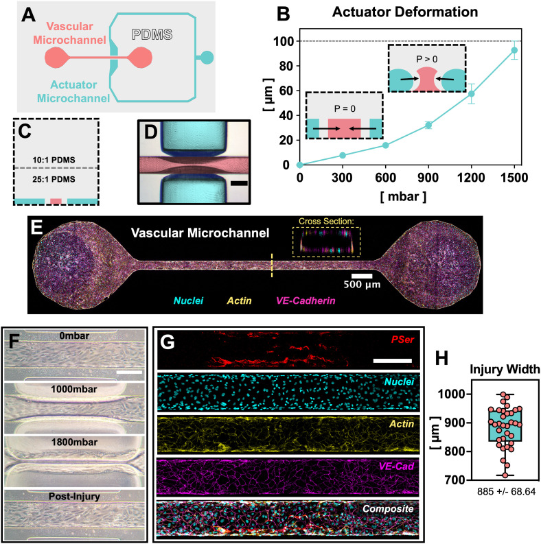
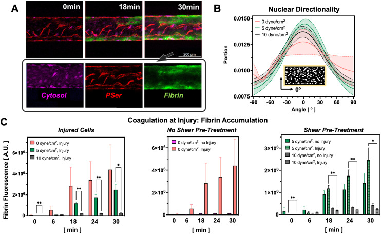
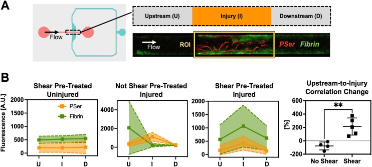
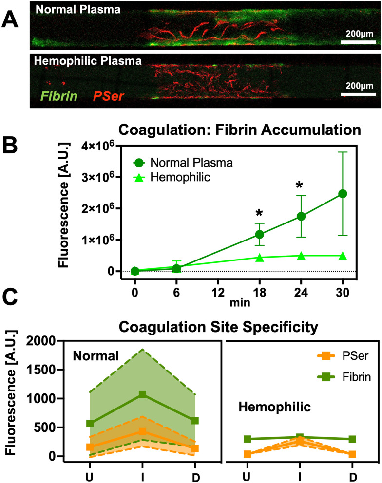
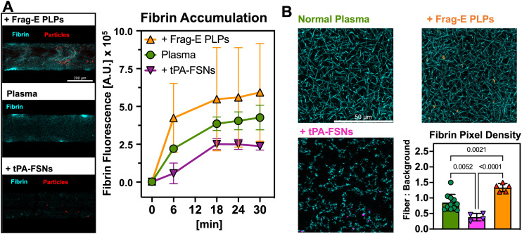
Blood coagulation is a highly regulated injury response that features polymerization of fibrin fibers to prevent the passage of blood from a damaged vascular endothelium. A growing body of research seeks to monitor coagulation in microfluidic systems but fails to capture coagulation as a response to disruption of the vascular endothelium. Here we present a device that allows compression injury of a defined segment of a microfluidic vascular endothelium and the assessment of coagulation at the injury site. This pressure injury-on-a-chip (PINCH) device allows visualization of coagulation as the accumulation of fluorescent fibrin at injury sites. Quantification of fluorescent fibrin levels upstream of and at injury sites confirm that pre-treating vascular endothelium with fluid shear stress helps capture coagulation as an injury response. We leverage the PINCH devices to demonstrate the limited coagulation response of type A hemophiliacs and evaluate the performance of hemostatic microparticles and fibrinolytic nanoparticles. Our findings and the straightforward fabrication of the PINCH devices make it a promising choice for additional screening of hemostatic therapeutics.


Conflict of interest statement

A. B. is a cofounder and stockholder of SelSym Biotech Inc. A. B. is an inventor on US Patent 10195304 and continuation in part 11419948B2 entitled “Functionalized microgels with fibrin binding elements”, licensed by SelSym.

Figures

Fig. 1
Fig. 1. A microvascular pressure injury-on-a-chip: “PINCH.” A) top-down schematic of microchannel design. A closed microchannel flanks a straight, vascular microchannel on two sides. B) When the actuator microchannel is pressurized, the PDMS walls between the actuator microchannel and vascular microchannel deform inward (mean ± SD, n = 6). For the 200 μm × 100 μm (w x h) vascular microchannel depicted here, actuator walls make initial contact at 1500 mbar. C) Deformation is enabled by curing a thin layer of soft, 25 : 1 PDMS immediately overtop microchannel molds. D) Deformation can be visualized via brightfield microscopy (scale: 200 μm). False colors are overlayed empty microchannels to indicate actuator and vascular microchannels. E) Confocal microscopy confirms staining for nuclei (cyan), F-actin (yellow) and VE-cadherin (magenta) via DAPI, phalloidin, and antibody staining, respectively. A patent lumen is confirmed by viewing the vascular microchannel cross section. F) Demonstration of HUVEC injury in a PINCH in phase contrast micrographs (scale: 200 μm). H) Confocal micrographs confirm antibody staining for the endothelial marker VE-cadherin (magenta) and annexin V staining for the cell death indicator phosphatidylserine (PSer, red). G) The width of injury regions is plotted as the apparent width of PSer-positive cells. Injury regions averaged 885 ± 68.64 μm (mean ± SD, n = 33).
Fig. 2
Fig. 2. Fluid shear stress and injury regulate coagulation levels in the PINCH devices. A) Confocal micrographs depicting accumulation of fluorescent fibrin (green) at PSer-positive (red) sites of vascular injury. Cytosolic dye (magenta) indicates uninjured cells. B) Nuclear directionality measurements reveal shear promotes more nuclear alignment along the direction of flow, 0° (n = 5). C) Pre-treatment of endothelium with fluid shear stress prior to injury limits subsequent coagulation (0 dyne cm−2 injury: N = 13; 0 dyne cm−2 no injury: N = 5; 5 dyne cm−2 injury: N = 6, 5 dyne cm−2 no injury: N = 5, 10 dyne cm−2 injury: N = 5, 10 dyne cm−2 no injury: N = 5; mean ± SEM, *p < 0.05, **p < 0.01, mixed-effects analysis). Greater coagulation levels are seen in injured vascular microchannels pre-treated with 0 and 5 dyne cm−2 shear stress as compared to 10 dyne cm−2.
Fig. 3
Fig. 3. Pre-treatment with extended fluid shear encourages injury site-specific coagulation during plasma perfusion. A) Fluorescent intensity of fibrin and phosphatidylserine was measured at, upstream, and downstream of injury sites. ROI were 900 μm wide and 240 μm tall. B) Fibrin intensity trends indicate that endothelium pre-treated with 5 dyne cm−2 fluidic shear stress are more efficient at capturing coagulation as an injury response compared to endothelium maintained at 0 dyne cm−2. Endothelium not pre-treated with fluidic shear stress are more likely to allow clotting upstream of injury sites. Pearson's correlation coefficient was calculated for fibrin and PSer. The percent change in PCC from upstream-to-injury depicts coagulation as being amplified by the injury. Correlation changes were compared with an unpaired, two-tailed Student's t-test **p = 0.0043. (Mean ± SD, n = 4–5).
Fig. 4
Fig. 4. Demonstration of reduced coagulation in hemophilic plasma. A) Confocal micrographs of vascular microchannel segments depict reduced coagulation of hemophilic plasma relative to controls. Although coagulation is reduced in hemophilic samples, an injury response is still visible as small regions of fibrin accumulation at the PSer-positive injury site. B and C) Quantification of fluorescent fibrin intensity reveal the coagulation response to injury is muted in hemophilic plasma. (Mean ± SD, n = 4–5, *p < 0.05, 2-way ANOVA).
Fig. 5
Fig. 5. Modification of coagulation with fibrin-binding microgels and nanogels. A) In the PINCH devices, coagulation levels of plasma supplemented with synthetic platelets confirm expected trends: Frag-E PLPs (n = 7) participate in coagulation by binding fibrin and collapsing via Brownian motion; tPA-FSNs (n = 3) disrupt coagulation by binding fibrin and releasing fibrinolytic tPA, halting the increase in coagulation seen in normal plasma (n = 4). (Mean ± SD). B) Confocal z-stack projections prepared on plain microscope slides confirm the trends seen in vascular microfluidics. The overall density of fibrin networks is increased or decreased by Frag-E PLPs and tPA-FSNs, respectively (mean ± SD, n = at least 3 slides, 1–3 images per slide; p-values displayed, one-way ANOVA).

References

    1. Yau J. W. Teoh H. Verma S. BMC Cardiovasc. Disord. 2015;15:1–11. doi: 10.1186/s12872-015-0124-z. - DOI - PMC - PubMed
    1. Nguyen T. C. and Carcillo J. A., in Pediatric Clinical Care, Springer, 2021, pp. 55–75
    1. Pradella P., Tomasella F. and Mascaretti L., in Hemocoagulative Problems in the Critically Ill Patient, Springer, 2012
    1. McRae S., in Mechanisms of Vascular Disease, Springer, 2020, pp. 199–213
    1. Alonso-Roman R. Mosig A. S. Figge M. T. Papenfort K. Eggeling C. Schacher F. H. Hube B. Gresnigt M. S. Nat. Microbiol. 2024;9:891–904. doi: 10.1038/s41564-024-01645-6. - DOI - PubMed

LinkOut - more resources