. 2024 Dec 23:14:1462231.
doi: 10.3389/fonc.2024.1462231. eCollection 2024.

Small-molecule MMRi36 induces apoptosis in p53-mutant lymphomas by targeting MDM2/MDM4/XIAP for degradation

Affiliations

Small-molecule MMRi36 induces apoptosis in p53-mutant lymphomas by targeting MDM2/MDM4/XIAP for degradation

Rati Lama et al. Front Oncol. .

Abstract

Rituximab combined with systemic chemotherapy significantly improves the rate of complete response in B-cell lymphomas. However, acquired rituximab resistance develops in most patients leading to relapse. The mechanisms underlying rituximab resistance are not well-understood. MDM2 and MDM4 proteins are major negative regulators of p53, but they also have p53-independent activities in mouse models of lymphomagenesis. Whether MDM2 or MDM4 is involved in rituximab resistance has not been explored. Here we report that MDM2 and MDM4 are upregulated in p53-mutant rituximab-resistant cells by transcriptional and post-transcriptional mechanisms. Knockdown of MDM2 or MDM4 significantly hindered growth of rituximab-resistant cells. To explore whether targeting the RING-domain of MDM2-MDM4 heterodimers is a viable strategy for the treatment of rituximab-resistant lymphomas, we identified MMRi36 in a high throughput small-molecule screen. Here we show that MMRi36 binds and stabilizes MDM2-MDM4 RING heterodimers and acts as an activator of the MDM2-MDM4 E3 ligase complex in vitro and promotes proteasomal degradation of MDM2/MDM4 proteins in cells. MMRi36 potently induces p53-independent apoptosis in p53-mutant lymphoma cells and it exerts non-apoptotic anti-lymphoma effect in rituximab resistant cells. The pro-apoptotic mechanisms of MMRi36 involves activation of both caspase 3 and caspase 7 associated with increased polyubiquitination and degradation of XIAP. Therefore, MMRi36 is a novel prototype small-molecule for targeting MDM2/MDM4/XIAP for degradation and induction of apoptosis in p53-mutant lymphomas.

Keywords: MDM2; MDM4; MMRi36; Rituximab; XIAP; apoptosis; lymphoma; mutant p53.


Conflict of interest statement

The authors declare that the research was conducted in the absence of any commercial or financial relationships that could be construed as a potential conflict of interest.

Figures

Figure 1
Figure 1
MDM4 and MDM2 play critical roles in proliferation of Rituximab-resistant cell lines (RRCL). (A) WB analysis of steady-state expression of MDM4, MDM2, p53 and Actin (loading control) proteins in parental and drug-resistant lymphoma cell lines. (B) qPCR analysis of MDM4 and MDM2 transcripts of the indicated cell lines. (C) Effect of MDM4 knockdown on growth of RL4RH cells in vitro, upper, WB of indicated proteins in sh control (shc) and two shMDM4 clones (shM4-1, shM4-2), lower, growth curves in a 4-day assay. Data were obtained from a single experiment with three duplicates. (D) Effect of MDM2 knockdown on growth of Raji4RH cells in vitro. The same as described in C except showing three shMDM2 clones. Data were obtained from a single experiment with three duplicates.
Figure 2
Figure 2
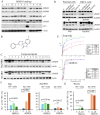
Identification of MMRi36 as potent apoptosis inducer in p53-mutant lymphoma and RRCL cells. (A) WB analysis of the indicated proteins in p53 (I254D)-mutant Ramos cells in a cell-based apoptosis inducer screen among analogs of primary hit MMRi3 of MDM2-MDM4 E3 ligase at 5 μM for 24h. aC3, activated caspase 3 and cleaved PARP (cPARP).C, non-treated control. (B) Chemical structure of MMRi36. (C) WB analysis of cPARP in RRCL cells after treatment with a panel of compounds at 5x IC50 concentrations for 24h (1, MMRi36 (5 μM), 2. Taxol (5 μM), 3, Doxorubicin (2 μM), 4, Carfilzomib (10 nM), 5, Vincristine (200 nM), 6, Etoposide (5 μM), 7, 5-Fluorouracil (100 μM), 8, Cytarabine (1 μM), 9, Carboplatin (20 μM), 10, Cisplatin (5 μM), 11, Entinostat (5 μM), 12, Bortezomib (20 nM), 13, Daunorubicin (5 μM), 14, MMRi64 (5 μM). Actin served as protein loading control. (D) WB analysis of apoptotic PARP cleavage in parental (RL, Raji) and RRCLs. (E) growth inhibition curves of indicated 4 cell lines in the presence of etoposide (upper) and MMRi36 (lower) and respective IC50s (right to the growth curves). Representative data of three independent experiments with three duplicates. (F, G) Quantification of apoptosis induced by MMRi36C in Raji and Raji4RH (F) or Ramos (G) cells. Cells were treated by MMRi36 at 5 and 10 μM for 24h followed by annexin-V-Alexa-488, PI and DAPI staining and image capture on Cytation 5. The annexin-V-positive only fractions (%) (AV+-only, early apoptotic cells) and annexin-V-positive/PI-positive (late-stage apoptotic cells) were shown. P values of unpaired student t test were shown, significant difference (*), very significant (**), or extremely significant (***). Representative data of two independent experiments with three duplicates.
Figure 3
Figure 3
MMRi36 binds to RING domain heterodimers and acts as an activator of the heterodimer MDM2-MDM4 E3 ligase in vitro. (A) In vitro E3 ubiquitin ligase assay with MDM2, MDM4 and p53 showing MMRi36 (10 μM) an activator while the primary hit MMRi3 (10 μM) an inhibitor of ubiquitination of MDM2-mediated p53 ubiquitination and polyubiquitination process. (B) In vitro pulldown assay for MMRi36 effect on RING-RING interaction of MDM2 and MDM4 with recombinant FLAG-MDM2B and MDM4 proteins. WB analysis of the FLAG-MDM4-bound MDM2B protein was shown. (C) MST assay using purified recombinant RING heterodimers of MDM2 and MDM to measure binding affinity of MMRi36 to the RING heterodimers. (D) Thermofluor (TF) assay with RING heterodimers and MMRi36, MMRi62 and solvent control (DMSO) showing MMRi36 stabilizes the heterodimers while MMRi62 destabilizes them. (E) In vitro ubiquitination assay showing concentration dependent effect of MMRi36 on E3 ligase activity of MDM2B-MDM4 toward MDM4, MDM2B and p53, and polyubiquitinated proteins.
Figure 4
Figure 4
MMRi36 induces MDM2/MDM4 downregulation in cells and induces p53–independent apoptosis in Caspase3/7-dependent manner. (A) WB analysis of indicated proteins in RL and RL4RH cells treated with 0 (C), 0.625, 1.25, 2.5, 5 and 10 μM of MMRi36 for 24h showing that MMRi36 downregulates MDM2 and MDM4 with activation of caspase 3 (AC3) and caspase 7 (AC7) and PARP cleavage (cPARP). The short isoform caspase 3 (sC3) band was shown. (B) WB analysis of apoptotic PARP cleavage in Raji4RH cells treated with 5 μM MMRi36 in the presence or absence of caspase3/7 inhibitor (C3/7i). (C) WB analysis of indicated proteins in indicated cells treated with either doxorubicin (25 nM) or MMRi36 (5 μM) for 24h.
Figure 5
Figure 5
MMRi36 downregulates XIAP by promoting XIAP polyubiquitination. (A) WB analysis of XIAP protein expression in RL and RL4RH cells treated with increasing concentrations of MMRi36 (0.63, 1.25, 2.5, 5, 10 μM) for 24h. (B) WB analysis of XIAP, PARP cleavage and activation of caspse3/7 in Raji4RH and Ramos with increasing concentrations of MMRi36 (1, 2, 4, 8 μM in Ramos cells and 2, 4, 8, 16 μM in Raji4RH cells) for 24h. AC7/AC3, activated caspase7/3. (C) WB analysis of XIAP protein expression in 293T cells transfected with XIAP alone or XIAP with MDM2B and MDM4 expression plasmids and treated with 5 μM MMRi62, MMRi67 or MMRi36 for 24h. (D) In vivo ubiquitination assay with the samples as in (C) in denatured His-ub pulldown followed by WB of XIAP showing increased polyubiquitinated XIAP after MMRi36 treatment.
Figure 6
Figure 6
A proposed model for MMRi36 induction of p53-independent apoptosis. MMRi36 independently targets the RING domains of MDM2-MDM4 (left cascade) and XIAP (right cascade) which activates their intrinsicE3 ligase activity toward themselves. Consequently, MMRi36 increases ubiquitination of MDM2, MDM4 and XIAP and their ubiquitin-dependent degradation in 26S proteasomes. Although low concentrations of MMRi36 increase p53 levels via downregulation of MDM2/MDM4, high concentrations MMRi36 promote ubiquitin-dependent degradation of p53. MMRi36-induced XIAP degradation is likely responsible for the p53-independent apoptosis induction by MMRi36.

Similar articles

Cited by

References

    1. Davis TA, Grillo-Lopez AJ, White CA, McLaughlin P, Czuczman MS, Link BK, et al. . Rituximab anti-CD20 monoclonal antibody therapy in non-Hodgkin's lymphoma: safety and efficacy of re-treatment. J Clin Oncol. (2000) 18:3135–43. doi: 10.1200/JCO.2000.18.17.3135 - DOI - PubMed
    1. Coiffier B, Lepage E, Briere J, Herbrecht R, Tilly H, Bouabdallah R, et al. . CHOP chemotherapy plus rituximab compared with CHOP alone in elderly patients with diffuse large-B-cell lymphoma. N Engl J Med. (2002) 346:235–42. doi: 10.1056/NEJMoa011795 - DOI - PubMed
    1. Habermann TM, Weller EA, Morrison VA, Gascoyne RD, Cassileth PA, Cohn JB, et al. . Rituximab-CHOP versus CHOP alone or with maintenance rituximab in older patients with diffuse large B-cell lymphoma. J Clin Oncol. (2006) 24:3121–7. doi: 10.1200/JCO.2005.05.1003 - DOI - PubMed
    1. Czuczman MS, Olejniczak S, Gowda A, Kotowski A, Binder A, Kaur H, et al. . Acquirement of rituximab resistance in lymphoma cell lines is associated with both global CD20 gene and protein down-regulation regulated at the pretranscriptional and posttranscriptional levels. Clin Cancer Res. (2008) 14:1561–70. doi: 10.1158/1078-0432.CCR-07-1254 - DOI - PubMed
    1. Olejniczak SH, Hernandez-Ilizaliturri FJ, Clements JL, Czuczman MS. Acquired resistance to rituximab is associated with chemotherapy resistance resulting from decreased Bax and Bak expression. Clin Cancer Res. (2008) 14:1550–60. doi: 10.1158/1078-0432.CCR-07-1255 - DOI - PubMed

LinkOut - more resources