. 2024 Dec 23:15:1521080.
doi: 10.3389/fimmu.2024.1521080. eCollection 2024.

Pseudomonas plecoglossicida infection induces neutrophil autophagy-driven NETosis in large yellow croaker Larimichthys crocea

Affiliations

Pseudomonas plecoglossicida infection induces neutrophil autophagy-driven NETosis in large yellow croaker Larimichthys crocea

Jia-Feng Cao et al. Front Immunol. .

Abstract

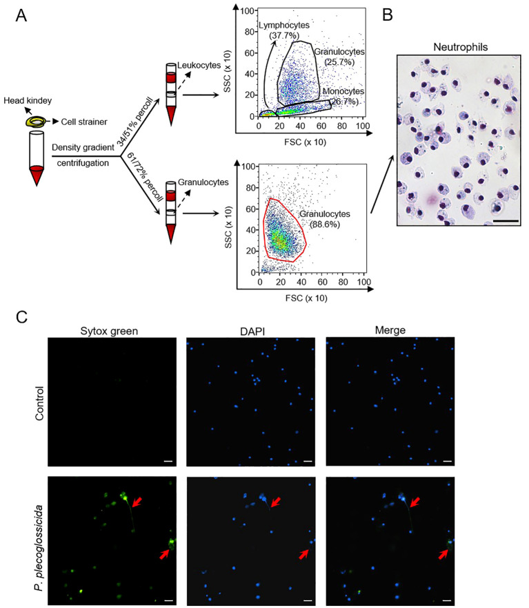
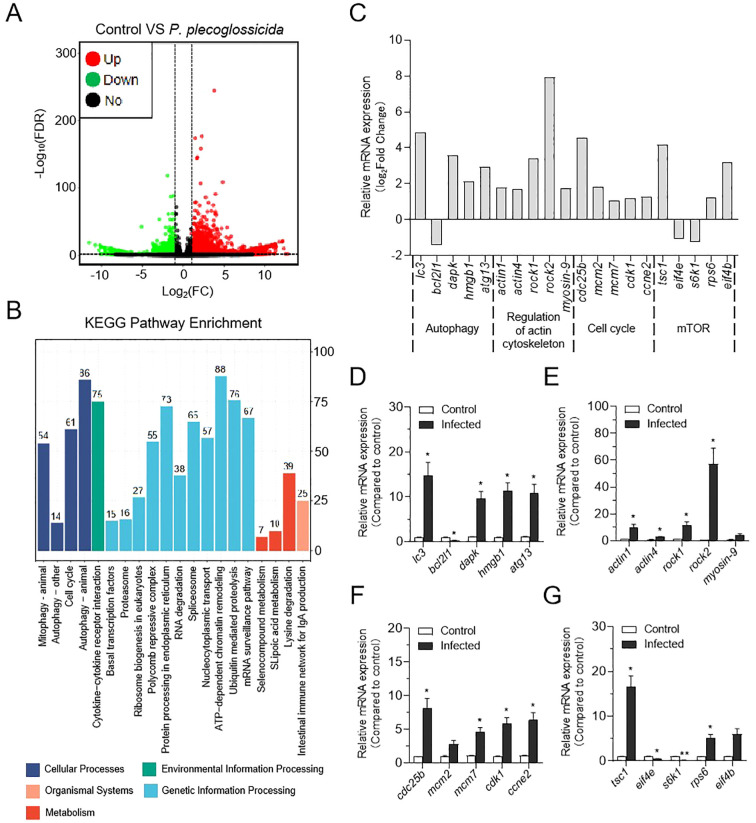
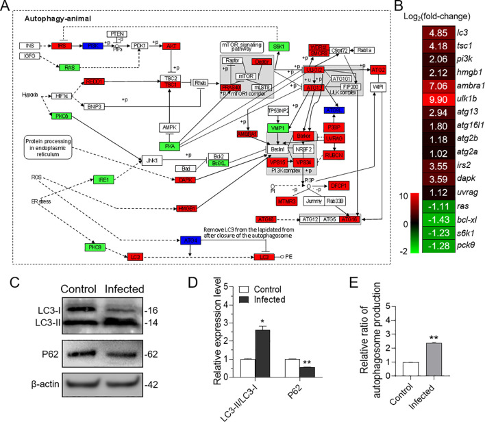
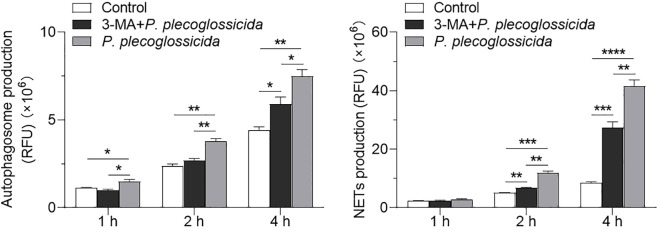
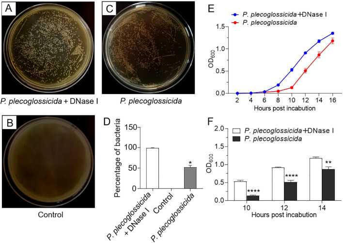
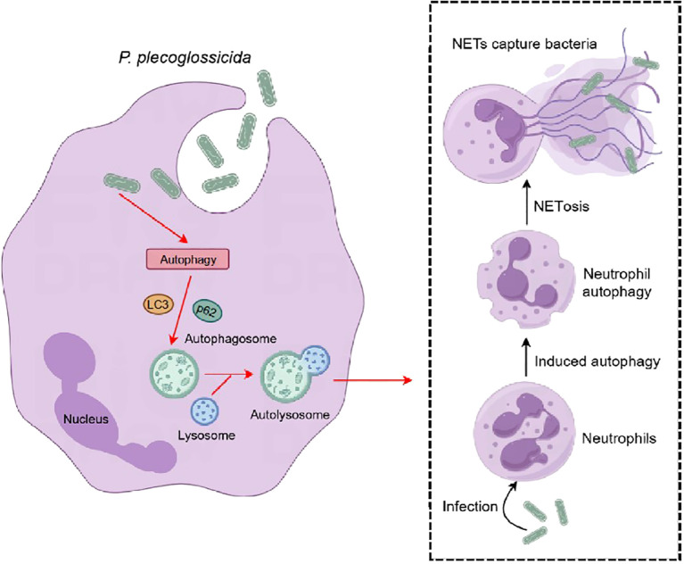
Neutrophil extracellular traps (NETs) are crucial for the immune defense of many organisms, serving as a potent mechanism for neutrophils to capture and eliminate extracellular pathogens. While NETosis and its antimicrobial mechanisms have been well studied in mammals, research on NETs formation in teleost fish remains limited. In this study, we used the large yellow croaker (Larimichthys crocea) as the study model to investigate NETosis and its role in pathogen defense. Our results showed that infection with Pseudomonas plecoglossicida could induce NETosis. To further explore the underlying mechanism, we performed transcriptome analysis and western blotting, which revealed that P. plecoglossicida triggers NETosis through activation of the autophagy pathway. Inhibition of autophagy significantly reduced NET production, highlighting its critical role in this process. Furthermore, our studies demonstrated that NETs exert a bacteriostatic effect, significantly suppressing the growth of P. plecoglossicida. Taken together, our findings reveal that autophagy regulates NETosis in large yellow croaker and underscore the essential role of NETs in bacterial defense, providing new insights into immune responses in teleost fish.

Keywords: Larimichthys crocea; Pseudomonas plecoglossicida; antibacterial; autophagy; neutrophil extracellular traps.


Conflict of interest statement

The authors declare that the research was conducted in the absence of any commercial or financial relationships that could be construed as a potential conflict of interest. The author(s) declared that they were an editorial board member of Frontiers, at the time of submission. This had no impact on the peer review process and the final decision.

Figures

Figure 1
Figure 1
Isolation and identification of large yellow croaker neutrophils and the effect of P. plecoglossicida on the NETs formation. (A) Isolation of leucocytes and neutrophils from the head kidney and validation by flow cytometry. (B) Giemsa staining of neutrophils. Scale bar, 20 μm. (C) Neutrophils in the control and P. plecoglossicida groups (P. plecoglossicida incubated with neutrophils for 2 h) were stained with SYTOX Green and DAPI and analyzed by immunofluorescence microscopy. Arrows indicate NETs, Scale bar, 20 μm.
Figure 2
Figure 2
Effect of P. plecoglossicida infection on transcriptome changes in neutrophils of the large yellow croaker. (A) Volcano plot showing the distribution of DEGs in the P. plecoglossicida infection group compared to the control group. (B) Counts of DEGs in each KEGG pathway. Different colors mean different classes. (C) The expression of major genes in the autophagy, regulatory actin cytoskeleton, cell cycle, and mTOR pathways in RNA sequencing. (D–G) qPCR validation of DEGs in the pathways of autophagy, regulation of the actin cytoskeleton, cell cycle, and mTOR (n = 3 per group). Statistical differences were performed by Student’s t-test. Data in (D–G) are representative of at least three independent experiments (Mean ± SEM). *P < 0.05, **P < 0.01.
Figure 3
Figure 3
Effect of in vitro infection of neutrophils with P. plecoglossicida on autophagy. (A) Both of red, green, and deep blue shading boxes represent molecules of the autophagy pathway identified in head kidney neutrophils of large yellow croaker. And the red boxes indicate the up-regulated DEGs in this pathway, the green boxes indicate the down-regulated DEGs in this pathway, and the deep blue boxes indicate both up-regulated and down-regulated DEGs in this pathway. (B) Differential expression genes involved in the autophagy pathway were analyzed after P. plecoglossicida infection. The color gradient represents highly up-regulated (red) to highly down-regulated (green) genes. (C) Immunoblot analysis of LC3 and p62/SQSTM1 protein levels in neutrophils infected with P. plecoglossicida for 2 h. (D) The relative proportions of LC3-II/LC3-I and p62/SQSTM1 in the infected compared with the control group were evaluated by densitometric analysis of immunoblots from (C) (n = 3 per group). (E) Neutrophils were infected with or without P. plecoglossicida for 2 h, and autophagy fluorescence intensity was detected (n = 3 per group). Statistical differences were performed by Student’s t-test. Data in (D, E) are representative of at least three independent experiments (Mean ± SEM). *P < 0.05, **P < 0.01.
Figure 4
Figure 4
Effect of the inhibition of autophagy on the NETs formation in neutrophils. (A) Large yellow croaker neutrophils were treated with or without 3-MA and then infected with P. plecoglossicida for different times to detect autophagy production. The control group was untreated (n = 3 per group). (B) Large yellow croaker neutrophils were treated with or without 3-MA and then infected with P. plecoglossicida for different times to detect NETs production. The control group was untreated (n = 3 per group). Statistical differences were performed by Student’s t-test. Data in (A, B) are representative of at least three independent experiments (Mean ± SEM). *P < 0.05, **P < 0.01, ***P < 0.001, ****P < 0.0001.
Figure 5
Figure 5
Effects of neutrophil NETs on survival and growth of P. plecoglossicida. (A–C) Large yellow croaker neutrophils were pretreated with PMA for 2h and then infected with P. plecoglossicida before being treated with or without DNase I for 6 h, and the collected mixtures (bacteria + neutrophils and NETs) were inoculated into the TSA agar plates to observe colony formation. The control group was untreated. (D) Graph showing the number of colonies in (A–C) (n = 3 per group). (E) Large yellow croaker neutrophils were infected with P. plecoglossicida and then treated with or without DNase I for 6 h, and the collected mixtures (bacteria + neutrophils and NETs) were inoculated into the TSB medium to observe the OD value of the P. plecoglossicida growth curve (n = 3 per group). (F) Statistical plots of OD values at 10, 12, and 14 h in F (n = 3 per group). Statistical differences were performed by Student’s t-test. Data in (D–F) are representative of at least three independent experiments (Mean ± SEM). *P < 0.05, **P < 0.01, ****P < 0.0001.
Figure 6
Figure 6
Pattern diagram of P. plecoglossicida induced NETosis in the large yellow croaker. After infection with P. plecoglossicida, the autophagy pathway is activated in large yellow croaker neutrophils, which in turn causes autophagy in neutrophils and induces NETosis, and the formation of NETs can capture the pathogenic bacterium P. plecoglossicida to prevent its invasion of the organism.

Similar articles

Cited by

References

    1. Mayadas TN, Cullere X, Lowell CA. The multifaceted functions of neutrophils. Annu Rev Pathol. (2014) 9:181–218. doi: 10.1146/annurev-pathol-020712-164023 - DOI - PMC - PubMed
    1. Chavakis E, Choi EY, Chavakis T. Novel aspects in the regulation of the leukocyte adhesion cascade. Thromb Haemost. (2009) 102:191–7. doi: 10.1160/TH08-12-0844 - DOI - PMC - PubMed
    1. Ley K, Laudanna C, Cybulsky MI, Nourshargh S. Getting to the site of inflammation: the leukocyte adhesion cascade updated. Nat Rev Immunol. (2007) 7:678–89. doi: 10.1038/nri2156 - DOI - PubMed
    1. Borregaard N. Neutrophils, from marrow to microbes. Immunity. (2010) 33:657–70. doi: 10.1016/j.immuni.2010.11.011 - DOI - PubMed
    1. Gazendam RP, van de Geer A, Roos D, van den Berg TK, Kuijpers TW. How neutrophils kill fungi. Immunol Rev. (2016) 273:299–311. doi: 10.1111/imr.12454 - DOI - PubMed

Supplementary concepts

LinkOut - more resources