Review
. 2024 Dec 23:15:1431112.
doi: 10.3389/fimmu.2024.1431112. eCollection 2024.

Metabolic adaptation of myeloid cells in the glioblastoma microenvironment

Affiliations
Review

Metabolic adaptation of myeloid cells in the glioblastoma microenvironment

Nora Essakhi et al. Front Immunol. .

Abstract

In recent decades, immunometabolism in cancers has emerged as an interesting target for treatment development. Indeed, the tumor microenvironment (TME) unique characteristics such as hypoxia and limitation of nutrients availability lead to a switch in metabolic pathways in both tumor and TME cells in order to support their adaptation and grow. Glioblastoma (GBM), the most frequent and aggressive primary brain tumor in adults, has been extensively studied in multiple aspects regarding its immune population, but research focused on immunometabolism remains limited. Here, we provide an overview of immunometabolism adaptation of myeloid cells in cancers with a specific focus on GBM and other brain tumors, before describing current therapeutic strategies targeting metabolic pathways. The main myeloid cells composing the GBM TME include tumor-associated macrophages (TAMs), which comprise both peripheral macrophages and local microglia, as well as myeloid-derived suppressor cells. The metabolic pathways involved in myeloid cell remodeling encompass the tricarboxylic acid cycle (TCA cycle), the lipid, glucose and amino acid metabolism and hypoxia. Developing treatments that target these metabolic pathways in tumor growth and its TME is a promising and increasing field. It includes both drug-repurposing and the development of innovative metabolic therapies. We finally provide an overview of all clinical trials in neuro-oncology involving treatments modifying cell metabolism and provide the preclinical rationale for both drugs already evaluated within clinical trials and potential candidates for future trials.

Keywords: TCA cycle; glioblastoma; glycolysis; lipid metabolism; metabolism; myeloid cells.


Conflict of interest statement

The authors declare that the research was conducted in the absence of any commercial or financial relationships that could be construed as a potential conflict of interest.

Figures
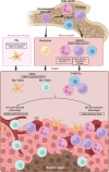
Figure 1
Figure 1
Myeloid cells lineage and location in GBM. Myeloid cells derive from bone marrow hematopoietic stem cells. They are consistently present in the blood circulation and can be recruited when needed. The main myeloid cells observed in the GBM microenvironment are the Tumor-infiltrating CD45+/CD33+/CD11b+ myeloid derived suppressor cells (MDSCs) as well as the CD45+/CD33+/HLA-DR+ tumor-associated macrophages (TAMs), including the local microglia (Mg-TAMs) and the peripheral macrophages/monocytes (Mo-TAMs). Secreted factors, hypoxia and lack of nutrients within the tumor microenvironment can influence MDSC and TAMs differentiation, directing them towards either a “M1” anti-tumoral phenotype or a “M2” pro-tumoral phenotype. While Mg-TAMs are mostly located in the periphery of the tumor, Mo-TAMs are found around the blood vessels and around the necrotic region. MDSCs are located near the cancer stem cells (CSC). The illustration was created with Biorender.com.
Figure 2
Figure 2
Schematic representation of the complex and heterogeneous immune microenvironment of GBM. (A) The Warburg effect describes the glycolysis dependence in hypoxic tumor cells. This process involves the co-activation of aurora kinase A (AURKA) with the proto-oncogene c-myc, leading to increased expression of lactate deshydrogenase A (LDHA) and hexokinase 2 (HK2). HK2 is essential in the first step of glycolysis, converting glucose into glucose-6-phosphate (G6P), which is further processed into phosphoenolpyruvate (PEP). PEP inhibits apoptosis by preventing excessive reactive oxygen species (ROS). PEP is then converted into pyruvate, and LDHA facilitates the transformation of pyruvate into lactate. This lactate is secreted into the tumor microenvironnement (TME), promoting M2 polarization, and the survival and proliferation of myeloid derived suppressor cells (MDSCs) and tumor-associated macrophages (TAMs). Additionally, HIF-1α inhibits oxidative phosphorylation (OXPHOS) metabolism. Cancer cells also secrete various factors that further support M2 polarization and the survival and proliferation of MDSCs and TAMs. (B) Factors secreted by cancer cells activate the JAK-STAT3/5 pathway in pro-tumoral “M2” myeloid derived suppressor cells (M2-MDSCs) and tumor-associated macrophages (TAMs). This activation increases the expression of fatty acid (FA) transporters such as Slc27a1, Slc27a6, Msr1 and CD36, leading to enhanced uptake of FAs and triacylglycerol, as well as increased lipid metabolism. FAs are used in FA oxidation (FAO) and the tricarboxylic acid (TCA) cycle, while triacylglycerols are absorbed into lysosomes for lipolysis. Metabolic modulation also includes increased acetyl-CoA acetyltransferase 1 (ACAT1) expression by M2-MDSCs/TAMs, which boost FAO and cholesterol metabolism. The enhanced TCA cycle activity raises cholesterol metabolism by transporting citrate out of the mitochondria for cholesterol synthesis. This upregulation in cholesterol metabolism leads to increased expression of immunosuppressors, such as Siglec-10 and PD-1. The anti-tumoral immune response is further inhibited by the LDHA secreted by tumor cells. This inhibition occurs through the higher expression of NKG2D ligands on the surface of M2-MDSCs/TAMs. By interacting with NKG2D receptor on NK cells surface, the ligands prevent the recognition of NKG2D ligand-bearing tumor cells and thus, impairs their anti-tumoral ability. Consequently, lipid metabolism is favored in M2-MDSCs/TAMs, with glycolysis also increased but to a lesser extent by SIRT1 and mTOR-HIF-1α pathway inhibition. Finally, M2-MDSCs/TAMs secrete the same factors as tumor cells, thereby auto-stimulating their own proliferation and infiltration. (C) The reverse Warburg effect corresponds to metabolic changes of cancer-associated fibroblasts (CAFs) due to oxidative tumor cells. An increase in reactive oxygen species (ROS) within these cells enhances glycolysis in CAFs, while reducing the expression of CAV-1. As a result, lactate is produced as byproduct of glycolysis and is secreted via the MCT4 transporter. Cancer cells then take up this lactate through MCT1 transporter to fuel oxidative phosphorylation (OXPHOS), which contributes to tumor proliferation. (D) In the tumor, both cancer cells and pro-tumoral immune cells increase their amino acid metabolism to survive and proliferate within the harsh tumor microenvironnement (TME). L-arginine is taken up by cancer cells and M2-MDSCs/TAMs and is subsequently degraded into ornithine and urea, leading to polyamines and glutamate. In tumor cells, arginase is upregulated, with Arg2 being more expressed than ArgI. The increased degradation of L-arginine into polyamines by ArgI plays a crucial role in tumor survival and proliferation, as well as in mTOR activation and glycolysis. In M2-MDSC/TAMs, ArgI is also upregulated, partly due to factors secreted by tumor cells, which also promote the recruitment and expansion of pro-tumoral cells. The resulting increase in polyamines raises the intracellular pH, allowing cells to survive in the low pH environment of the TME induced by lactate. ArgI is also secreted by these cells, further depleting the TME of L-arginine. This depletion impairs CD3ζ/CD3ϵ receptors on T cells, hindering their function. Another amino acid depletion in the TME that leads to immunosuppression is the Glutamine. The Glutamine pathway is highly active in both tumor and pro-tumoral cells, with a high uptake of Glutamine through SLC7A5 transporter in cancer cells and mGluR2/3 in immune cells. In cancer cells, Glutamine is used for protein synthesis or converted into Glutamate. Glutamate can be used in the TCA cycle and FAO, or to activate the mTOR/glycolysis pathway and the FA synthesis. The steps leading to α-KG production from Glutamine release NH4+, which accumulates and promotes autophagy, aiding tumor survival and proliferation. Tumor cells also secrete some of the produced Glutamate, which is then recaptured by pro-tumoral immune cells. The increase in Glutamate within the cells is due to heightened Glutamine uptake, increased L-arginine metabolism, and secretion by cancer cells. As in cancer cells, Glutamate in immune cells is used to produce α-KG for FA synthesis, participate in the TCA cycle and FAO, and activate mTOR and glycolysis. This latter pathway is also activated by the high L-arginine uptake. The illustration was created with Biorender.com.

References

    1. Louis DN, Perry A, Wesseling P, Brat DJ, Cree IA, Figarella-Branger D, et al. . The 2021 WHO classification of tumors of the central nervous system: a summary. Neuro-Oncology. (2021) 23:1231–51. doi: 10.1093/neuonc/noab106 - DOI - PMC - PubMed
    1. Stupp R, Mason WP, van den Bent MJ, Weller M, Fisher B, Taphoorn MJB, et al. . Radiotherapy plus concomitant and adjuvant temozolomide for glioblastoma. New Engl J Med. (2005) 352:987–96. doi: 10.1056/NEJMoa043330 - DOI - PubMed
    1. Jung E, Osswald M, Ratliff M, Dogan H, Xie R, Weil S, et al. . Tumor cell plasticity, heterogeneity, and resistance in crucial microenvironmental niches in glioma. Nat Commun. (2021) 12:1014. doi: 10.1038/s41467-021-21117-3 - DOI - PMC - PubMed
    1. Broekman ML, Maas SLN, Abels ER, Mempel TR, Krichevsky AM, Breakefield XO. Multidimensional communication in the microenvirons of glioblastoma. Nat Rev Neurol. (2018) 14:482–95. doi: 10.1038/s41582-018-0025-8 - DOI - PMC - PubMed
    1. Guo C, Chen S, Liu W, Ma Y, Li J, Fisher PB, et al. . Immunometabolism: A new target for improving cancer immunotherapy. Adv Cancer Res. (2019) 143:195–253. doi: 10.1016/bs.acr.2019.03.004 - DOI - PMC - PubMed

MeSH terms

LinkOut - more resources