[Preprint]. 2025 Apr 14:2024.12.16.628822.
doi: 10.1101/2024.12.16.628822.

De novo design and structure of a peptide-centric TCR mimic binding module

Affiliations

De novo design and structure of a peptide-centric TCR mimic binding module

Karsten D Householder et al. bioRxiv. .

Update in

Abstract

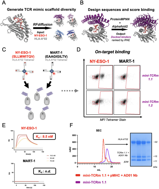
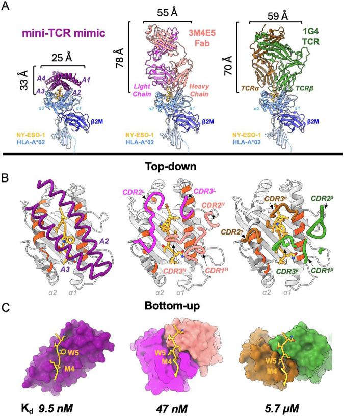
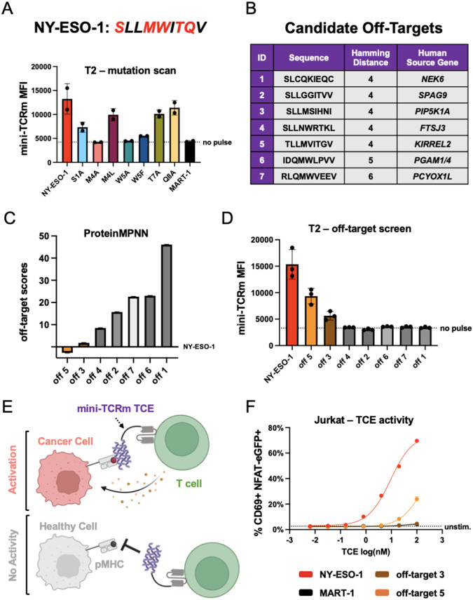
T cell receptor (TCR) mimics offer a promising platform for tumor-specific targeting of peptide-MHC in cancer immunotherapy. Here, we designed a de novo α-helical TCR mimic (TCRm) specific for the NY-ESO-1 peptide presented by HLA-A*02, achieving high on-target specificity with nanomolar affinity (Kd = 9.5 nM). The structure of the TCRm/pMHC complex at 2.05 A resolution revealed a rigid TCR-like docking mode with an unusual degree of focus on the up-facing NY-ESO-1 side chains, suggesting the potential for reduced off-target reactivity. Indeed, a structure-informed in silico screen of 14,363 HLA-A*02 peptides correctly predicted two off-target peptides, yet our TCRm maintained a wide therapeutic window as a T cell engager. These results represent a path for precision targeting of tumor antigens with peptide-focused α-helical TCR mimics.


Figures

Fig. 1.
Fig. 1.. Design and experimental validation of de novo mini-TCR mimics.
(A) Schematic of RFdiffusion fold-conditioning pipeline to generate four scaffolds over the NY-ESO-1 HLA-A*02 input (red and gray). (B) Schematic of pipeline using ProteinMPNN to design sequences for Scaffold #1 (purple) and predict binders with AlphaFold2. (C) Yeast display screening of top de novo designs with NY-ESO-1 (red) vs. MART-1 HLA-A*02 (black) tetramers. (D) Binders staining NY-ESO-1 but not MART-1 HLA-A*02 tetramer by flow cytometry. (E) SPR analysis of mini-TCRm 1.1 analyte with immobilized NY-ESO-1 HLA-A*02 (red) vs. MART-1 HLA-A*02 (black). Dissociation constants indicated on sensograms (Kd ); n.d., not determined; RU, response unit. (F) SEC profiles of mini-TCRm 1.1 (purple) vs. co-eluting complex (red) of mini-TCRm 1.1, NY-ESO-1 HLA-A*02, and AD01 nanobody. SDS-Page gel showing fractions of co-eluting complex pooled for crystal screens. mAU, milli-Absorbance Units; kDa, kilodaltons.
Fig. 2.
Fig. 2.. High-resolution crystal structure reveals peptide-specific interactions.
(A) Front view of mini-TCRm/pMHC complex (PDB ID 9MIN). A1-A4 alpha-helices of mini-TCRm (purple); α1 and α2 helices of HLA-A*02 and full chain (light blue); NY-ESO-1 peptide (gold); β2M (dark blue); AD01 nanobody (white). (B) Side view of complex with Met4-Trp5 bulge on the NY-ESO-1 peptide. (C) Top-down view of complex. (D) Bottom-up view of peptide-specific interactions with surface of mini-TCRm (purple). Box zooming in on NY-ESO-1’s Met4-Trp5 motif (gold) fitting into mini-TCRm pockets formed between the A2 and A3 helices (key residue labels in white). (E) Hydrogen bonds (dashed lines) and key peptide-centric residues between mini-TCRm and NY-ESO-1 peptide. (F) Hydrogen bonds and salt bridges (dashed lines) between mini-TCRm and HLA-A*02.
Fig. 3.
Fig. 3.. Comparison to existing antibody TCR mimic and natural TCR.
(A) Front view comparison between mini-TCRm (purple), 3M4E5 Fab (magenta and salmon), and 1G4 TCR (brown and green) bound to NY-ESO-1 peptide (gold) and HLA-A*02 (light blue). Height and width of binders shown in Angstroms (Å). (B) Top-down comparison of docking footprints of each binder with MHC contact residues (red orange) and NY-ESO-1 peptide side chains (gold) (C) Bottom-up comparison of interactions between the NY-ESO-1 peptide (gold) and binding pockets, with the Met4-Trp5 motif labeled. Dissociation constants (Kd) for each complex is shown at the bottom. nanomolar, nM; micromolar, μM.
Fig. 4.
Fig. 4.. Structure-guided identification of off-target peptides reveals a robust therapeutic window.
(A) Mutation scanning the NY-ESO-1 peptide (sequence shown above plot; mutated residues in red) and pulsing on T2 cells. Variants are named by original residue, position, followed by the mutation. Measured mini-TCRm MFI (Mean Fluorescence Intensity) by flow cytometry. (B) Table of candidate off-targets from the MHC Motif Atlas. (C) Ranking of candidate off-targets based on ProteinMPNN and the crystal structure. (D) Pulsing candidate off-target peptides on T2 cells and staining with mini-TCRm. (E) Schematic of a mini-TCRm T cell engager (TCE; purple) activating T cells against a cancer cell (salmon) with an on-target peptide (red) but not a healthy cell (gray) with an off-target peptide (black). (F) T2-Jurkat co-culture assay with pulsed peptides and mini-TCRm TCE. T cell activation shown as percentage of CD69+ NFAT-eGFP+ Jurkat cells versus no-pulse control from flow cytometry analysis. TCE dose shown in nanomolar on the log scale (log(nM)).

References

    1. Xie N., Shen G., Gao W., Huang Z., Huang C., Fu L., Neoantigens: promising targets for cancer therapy. Sig Transduct Target Ther 8, 9 (2023). - PMC - PubMed
    1. Klebanoff C. A., Chandran S. S., Baker B. M., Quezada S. A., Ribas A., T cell receptor therapeutics: immunological targeting of the intracellular cancer proteome. Nat Rev Drug Discov 22, 996–1017 (2023). - PMC - PubMed
    1. Rossjohn J., Gras S., Miles J. J., Turner S. J., Godfrey D. I., McCluskey J., T Cell Antigen Receptor Recognition of Antigen-Presenting Molecules. Annu. Rev. Immunol. 33, 169–200 (2015). - PubMed
    1. Cresswell P., Ackerman A. L., Giodini A., Peaper D. R., Wearsch P. A., Mechanisms of MHC class I-restricted antigen processing and cross-presentation. Immunological Reviews 207, 145–157 (2005). - PubMed
    1. Leko V., Rosenberg S. A., Identifying and Targeting Human Tumor Antigens for T Cell-Based Immunotherapy of Solid Tumors. Cancer Cell 38, 454–472 (2020). - PMC - PubMed

Publication types