[Preprint]. 2024 Dec 18:2024.12.17.628992.
doi: 10.1101/2024.12.17.628992.

The pseudoknot structure of a viral RNA reveals a conserved mechanism for programmed exoribonuclease resistance

Affiliations

The pseudoknot structure of a viral RNA reveals a conserved mechanism for programmed exoribonuclease resistance

Jeanine G Gezelle et al. bioRxiv. .

Abstract

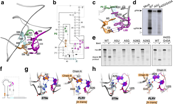
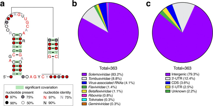
Exoribonuclease-resistant RNAs (xrRNAs) are viral RNA structures that block degradation by cellular 5'-3' exoribonucleases to produce subgenomic viral RNAs during infection. Initially discovered in flaviviruses, xrRNAs have since been identified in wide range of RNA viruses, including those that infect plants. High sequence variability among viral xrRNAs raises questions about the shared molecular features that characterize this functional RNA class. Here, we present the first structure of a plant-virus xrRNA in its active exoribonuclease-resistant conformation. The xrRNA forms a 9 base pair pseudoknot that creates a knot-like topology similar to that of flavivirus xrRNAs, despite lacking sequence similarity. Biophysical assays confirm a compact pseudoknot structure in solution, and functional studies validate its relevance both in vitro and during infection. Our study reveals how viral RNAs achieve a common functional outcome through highly divergent sequences and identifies the knot-like topology as a defining feature of xrRNAs.


Figures

Figure 1.
Figure 1.. A compact RNA structure in the 3′ UTR of the tombusvirus-associated RNA ST9a blocks degradation by Xrn1.
a) Northern blot of total RNA from N. benthamiana mock-infected or infected with ST9a for 8 days. Probes are against the ST9a 3′ UTR. The white arrow denotes an unspecified RNA species, likely due to additional in-cell processing of the sgRNA or host rRNA trapping. b) Sequencing gel of cDNA from Xrn1 degradation reaction (1), crystallization construct (2) and undigested RNA (3) to map the Xrn1 stop site at single nucleotide resolution. Dideoxy sequencing lanes are labeled and used to determine the sequence around the Xrn1 stop site, depicted on the right with arrows. c) In vitro Xrn1 degradation reaction of ST9a 3′ UTR to determine the minimal Xrn1-resistant element. Reactions were resolved by dPAGE and visualized by ethidium bromide staining. Length in nucleotides (nt) corresponds to the resistant product, counting from the Xrn1 stop site as determined in b. d) Secondary structure diagram of the ST9a xrRNA crystallization construct. Non–Watson–Crick base pairs are in Leontis–Westhof annotation and the Xrn1 stop site is depicted by the arrow. Non-modeled nucleotides are shown as lowercase letters. e) Ribbon diagram of the ST9a xrRNA crystal structure. Colors match d.
Figure 2.
Figure 2.. The structure of the ST9a xrRNA contains a 9 base pair PK and a protective ring around the 5′ end.
a) Ribbon diagram (right) and 2D structure diagram (left) of the ST9a xrRNA structure with the 9 base pair PK in blue, the 5′ end in cyan and the protective ring around the 5′ end in purple. b) Coaxial stacking of the PK and L2B region. Colors match Fig. 1d–e. c) In vitro Xrn1 degradation reaction of ST9a xrRNA wild type (WT) RNA and the indicated mutants. Reactions were resolved by dPAGE and visualized by ethidium bromide staining. d) Northern blot of total RNA from N. benthamiana mock-infected or infected with ST9a WT or the indicated mutants. Probes are against the ST9a 3′ UTR. The white arrow denotes an unspecified RNA species, likely due to additional in-cell processing of the sgRNA or host rRNA trapping. e) In vitro Xrn1 degradation reaction of ST9a xrRNA WT RNA and the indicated mutants. Reactions were resolved by dPAGE and visualized by ethidium bromide staining. f) Details of the interactions between A12 and A13 with the minor groove of the PK.
Figure 3.
Figure 3.. A conserved interaction network forms the core of the xrRNA structure.
a) Details of the tertiary interaction network centered around the L2B region with colors to match Fig. 1d–e. b) 2D diagram of the tertiary interaction network depicted in a. Non–Watson–Crick base pairs are in Leontis–Westhof annotation. c) Details of the L2A pi-stacking network with colors to match a. d) Northern blot of total RNA from N. benthamiana mock-infected or infected with ST9a WT or the indicated mutants. Probes are against the ST9a 3′ UTR. The white arrow denotes an unspecified RNA species, likely due to additional in-cell processing of the sgRNA or host rRNA trapping. e) In vitro Xrn1 degradation reaction of ST9a xrRNA WT RNA and the indicated mutants. Reactions were resolved by dPAGE and visualized by ethidium bromide staining. f-h) Conserved base triple interactions, schematically depicted in (f), centered around A5 (g) and A29 (h) are part of the conserved ST9a xrRNA core. Identical hydrogen bonds are formed in cis in ST9a xrRNA (left) and in trans between 2 RNA molecules in PLRV xrRNA (right, PDB: 7JJU).
Figure 4.
Figure 4.. Coordinated metal ions stabilize the xrRNA structure.
a) Quantitative Xrn1 degradation assay with 3′−32P-labeled ST9a xrRNA at varying Mg2+ concentrations. n=4. Error bars show SD. **** adjusted P-value <0.0001. b) Thermal melting curves of the ST9a xrRNA at the indicated Mg2+ concentrations. Shown is the first derivative of melting curves at 266 nm. Samples were measured in biological replicates (n=3). c) Iridium(III) hexammine ions in the major groove of the L2B-PK helix. d) Details of a conserved iridium(III) hexammine binding site in the ST9a xrRNA structure (left) and PLRV xrRNA structure (right). Note that the iridium(III) hexammine ion is coordinated by nucleotides from two RNA molecules of the crystallographic dimer in the PLRV xrRNA. e) Zoom-in to SEC profiles of the indicated ST9a xrRNA constructs at 0 or 10 mM Mg2+. f) Plot of Rg and g) scattering-pair distance distribution (P(r)) profiles derived from SAXS data for the monomeric peaks from e, showing Mg2+ induced compaction. See also Extended Data Figures 6 and 7 and Table 2.
Figure 5.
Figure 5.. Widespread distribution of class 3 xrRNAs in plant-infecting RNA viruses.
a) Consensus sequence secondary structure of class 3 xrRNAs based on a comparative sequence alignment of 363 sequences of plant-infecting RNA viruses and virus-associated RNAs. Y = pyrimidine; R = purine. b) Distribution of xrRNA sequences from (a) across viral genera. Note that many xrRNA-containing viruses previously annotated as Luteoviridae, have been re-classified as Solemoviridae. c) Metagenome analysis of xrRNA sequences from (a).
Figure 6.
Figure 6.. Structural comparison of class 1 and class 3 xrRNAs uncovers a conserved molecular mechanism to block host exoribonucleases.
a) 2D structure of the ST9a (left) and ZIKV (right) xrRNAs. Colors indicate common features. Lowercase letters in ZIKV xrRNA denote sequences that were mutated to aid crystallization. b) Overlay of the core of ST9a and ZIKV xrRNA with colors to match a. c) The protective rings of ST9a (top) and ZIKV (bottom) xrRNAs have similar dimensions. Colors to match a. The cyan sphere indicates the position of 5′ end. d) Details of the nucleotides positioning the 5′ end within the center of the ring of ST9a (top) and ZIKV (bottom) xrRNA. e) Details of the helix buttressing the 5′ side of the protective ring in ST9a (top) and ZIKV (bottom) xrRNA. f) Details of the unique interaction networks stabilizing the 3′ side of the protective ring in ST9a (top) and ZIKV (bottom) xrRNA.

References

    1. Pierson T. C. & Diamond M. S. The continued threat of emerging flaviviruses. Nat Microbiol 5, 796–812 (2020). - PMC - PubMed
    1. Keusch G. T. et al. Pandemic origins and a One Health approach to preparedness and prevention: Solutions based on SARS-CoV-2 and other RNA viruses. Proceedings of the National Academy of Sciences 119, e2202871119 (2022). - PMC - PubMed
    1. Jaafar Z. A. & Kieft J. S. Viral RNA structure-based strategies to manipulate translation. Nat Rev Microbiol 17, 110–123 (2019). - PMC - PubMed
    1. Smyth R. P., Negroni M., Lever A. M., Mak J. & Kenyon J. C. RNA Structure—A Neglected Puppet Master for the Evolution of Virus and Host Immunity. Front. Immunol. 9, (2018). - PMC - PubMed
    1. Falese J. P., Donlic A. & Hargrove A. E. Targeting RNA with small molecules: from fundamental principles towards the clinic. Chem Soc Rev 50, 2224–2243 (2021). - PMC - PubMed

Publication types

LinkOut - more resources