[Preprint]. 2024 Dec 17:2024.12.17.628915.
doi: 10.1101/2024.12.17.628915.

METTL7A improves bovine IVF embryo competence by attenuating oxidative stress

Affiliations

METTL7A improves bovine IVF embryo competence by attenuating oxidative stress

Linkai Zhu et al. bioRxiv. .

Update in

Abstract

In vitro fertilization (IVF) is a widely used assisted reproductive technology to achieve a successful pregnancy. However, the acquisition of oxidative stress in embryo in vitro culture impairs its competence. Here, we demonstrated that a nuclear coding gene, methyltransferase-like protein 7A (METTL7A), improves the developmental potential of bovine embryos. We found that exogenous METTL7A modulates expression of genes involved in embryonic cell mitochondrial pathways and promotes trophectoderm development. Surprisingly, we discovered that METTL7A alleviates mitochondrial stress and DNA damage and promotes cell cycle progression during embryo cleavage. In summary, we have identified a novel mitochondria stress eliminating mechanism regulated by METTL7A that occurs during the acquisition of oxidative stress in embryo in vitro culture. This discovery lays the groundwork for the development of METTL7A as a promising therapeutic target for IVF embryo competence.

Keywords: DNA damage; IVF; METTL7A; embryo competence; mitochondria; oxidative stress.


Conflict of interest statement

Competing interests The findings of this study were included in a U.S provisional patent application 63/698,174.

Figures
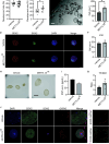
Figure 1.
Figure 1.. Exogenous METTL7A improves the developmental potential of bovine IVP embryos.
A. The cleavage OF METTL7A OE embryos (n = 29) compared to the vehicle control (n = 18). B. The blastocyst rate of METTL7A OE embryos (n = 11) compared to the control (n = 11). C. A representative image of bovine METTL7A OE embryos, scale bar = 100 μm. D. Immunostaining analysis of CDX2 (trophectoderm, TE marker) and SOX2 (inner cell mass, ICM marker) in METTL7A OE embryos compared to the control, scale bar = 50μm. E. The total TE cell number counts between METTL7A OE blastocysts (n = 5) compared to the control (n = 5). F. The total ICM cell number counts between METTL7A OE blastocysts (n = 5) compared to the control (n = 5). G. The TE/ICM cell number ratio between METTL7A OE blastocysts compared to the control. H. A representative bright field image of day 12 (E12) elongated embryos from METTL7A OE and control embryo transfer, scale bar = 500 μm. I. The serum INF-tau levels of surrogate cows with METTL7A OE embryo transfer (n = 2) compared to the control (n = 2) on the day of flushing. J. Immunostaining analysis of CDX2 (TE marker), SOX2 (embryonic disc marker), and GATA6 (hypoblast marker) in E12 embryos flushed out from the METTL7A OE and control embryo transfer, scale bar = 50μm.
Figure 2.
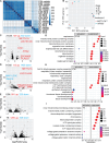
Figure 2.. The transcriptomic analysis of METTL7A OE embryos at 2-, 8-cell, and ICM/TE compared to control.
A. Heatmap of the samples from the same stages of bovine embryos from the METTL7A OE and control group. B. Principal component analysis (PCA) of the transcriptomes of METTL7A OE and control embryos at different developmental stages. C-F Volcano plots showing the number of up- or down-regulated genes in METTL7A OE embryos compared to control at 2-cell (C), 8-cell (D), ICM (E) and TE (F) stages (FDR < 0.05, |log2FC| > 1). The most significant up-regulated genes in METTL7A OE embryos compared to the control are highlighted. (G-I) The top GO terms of up- and down-regulated genes METTL7A OE embryos compared to the control at of 8-cell (G), ICM (H) and TE (I) stages.
Figure 3.
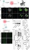
Figure 3.. Exogenous METTL7A reduces mitochondrial stress and decreases superoxide level of bovine pre-implantation embryos.
A. Experimental scheme of injecting METTL7A IVT-RNA into one blastomere of 2-cell embryos. B. Immunostaining analysis of F-actin, mitochondrial stress marker (HIF1a), and METTL7A-6xHisTag in METTL7A OE blastomeres and control. The red arrow points to condensed chromatin and degradation of actin filament, scale bar = 50μm. After 12 hours of injection, METTL7A OE blastomeres developed through 1 or 2 cell cycles, while non-injected blastomeres were arrested (n = 5, 4/5 or 80%) with described phenotype. C. Experimental scheme of zygotic injection and western blot analysis. D and E. Western blot analysis of Succinate Dehydrogenase Complex Flavoprotein Subunit A (SDHA, a marker for mitochondrial respiratory activity) in METTL7A OE (n = 6) blastocysts compared to control (n = 7). F. The immunostaining analysis of superoxide level measured by MitoSox green in METTL7A OE embryos and control at 8-cell (n = 13 embryos for both groups) and blastocyst (n = 10 embryos for both groups) stage, scale bar, 50μm. G and H. The quantification of superoxide level in METTL7A OE embryos and control at 8-cell (G) and blastocyst stage (H). I and J. The GSH level METTL7A OE embryos and control at 8-cell (I, n = 6 for both groups) and blastocyst stage (J, n = 3 for both groups).
Figure 4.
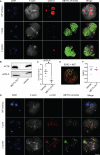
Figure 4.. Exogenous METTL7A attenuates DNA damage and promotes cell cycle progression.
A. Immunostaining analysis of F-actin, yH2A.X (DNA damage marker) and METTL7A-6xHisTag in METTL7A OE blastomeres (n = 5) and control (non-injected blastomeres and IVF embryos: METTL7A OE negative blastomere), scale bar = 50μm. B and C. Western blot analysis and quantification of yH2A.X in METTL7A OE embryos (n = 3) and the vehicle control (n = 4) at blastocyst stage 7 days after zygotic injection. D. Immunostaining analysis of p-Chk1 (cell cycle checkpoint marker) in METTL7A OE and IVF control embryos at the cleavage stage, scale bar = 50μm. E and F. Immunostaining analysis of the proliferative cells (Ki67+) in METTL7A OE blastocyst and control group. After 12 hours of zygotic injection, METTL7A OE blastomeres developed through 1 or 2 cell cycles, while non-injected blastomeres were arrested (n = 5).

Similar articles

Cited by

References

    1. 2021 Assited Reproduction Technology (ART) Fertility Clinic and National Summary Report [https://www.cdc.gov/art/reports/2021/summary.html]
    1. Viana JHM: 2022 Statistics of embryo production and transfer in domestic farm animals: the main trends for the world embryo industry still stand. In: Embryo Technology Newsletter. vol. 41. International Embryo Technologies Society; 2023: 20–38.
    1. Gardner DK, Kelley RL: Impact of the IVF laboratory environment on human preimplantation embryo phenotype. J Dev Orig Health Dis 2017, 8(4):418–435. - PubMed
    1. Hansen PJ: Implications of Assisted Reproductive Technologies for Pregnancy Outcomes in Mammals. Annu Rev Anim Biosci 2020, 8:395–413. - PubMed
    1. Lin J, Wang L: Oxidative Stress in Oocytes and Embryo Development: Implications for In Vitro Systems. Antioxid Redox Signal 2020. - PubMed

Publication types

LinkOut - more resources