This is a preprint.
Cell type transcriptional identities are maintained in cultured ex vivo human brain tissue
- PMID: 39763930
- PMCID: PMC11702615
- DOI: 10.1101/2024.12.19.629223
Cell type transcriptional identities are maintained in cultured ex vivo human brain tissue
Abstract
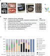
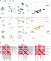
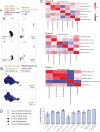
It is becoming more broadly accepted that human-based models are needed to better understand the complexities of the human nervous system and its diseases. The recently developed human brain organotypic culture model is one highly promising model that requires the involvement of neurosurgeons and neurosurgical patients. Studies have investigated the electrophysiological properties of neurons in such ex vivo human tissues, but the maintenance of other cell types within explanted brain remains largely unknown. Here, using single-nucleus RNA sequencing, we systematically evaluate the transcriptional identities of the various cell types found in six patient samples after fourteen days in culture (83,501 nuclei from day 0 samples and 45,738 nuclei from day 14 samples). We used two pediatric temporal lobectomy samples, an adult frontal cortex sample, two IDH wild-type glioblastoma samples, and one medulloblastoma sample. We found remarkably high correlations of day 14 transcriptional identities to day 0 tissue, especially in tumor cells (r = 0.90 to 0.93), though microglia (r = 0.86), oligodendrocytes (r = 0.80), pericytes (r = 0.77), endothelial cells (r = 0.78), and fibroblasts (r = 0.76) showed strong preservation of their transcriptional profiles as well. Astrocytes and excitatory neurons showed more moderate preservation (r = 0.66 and 0.47, respectively). Because the main difficulty with organotypic brain cultures is the acquisition of human tissue, which is readily available to neurosurgeons, this model is easily accessible to neurosurgeon-scientists and neurosurgeons affiliated with research laboratories. Broad uptake of this more representative model should prompt advances in our understanding of many uniquely human diseases, lead to more reliable clinical trial performance, and ultimately yield better therapies for our patients.
Keywords: Human brain; brain model development; epilepsy; glioblastoma; human brain organotypic slice culture; medulloblastoma.
Conflict of interest statement
Declaration of interest statement None of the authors have any conflict of interest with the study or any of the topics discussed.
Figures


References
-
- He Z, Dony L, Fleck JS, Szałata A, Li KX, Slišković I, Lin HC, Santel M, Atamian A, Quadrato G, Sun J, Pașca SP; Human Cell Atlas Organoid Biological Network; Camp JG, Theis FJ, Treutlein B. An integrated transcriptomic cell atlas of human neural organoids. Nature. 2024. Nov;635(8039):690–698. doi: 10.1038/s41586-024-08172-8. Epub 2024 Nov 20. Erratum in: Nature. 2024 Dec 11. - DOI - PMC - PubMed
-
- Chaichana KL, Capilla-Gonzalez V, Gonzalez-Perez O, Pradilla G, Han J, Olivi A, Brem H, Garcia-Verdugo JM, Quiñones-Hinojosa A. Preservation of glial cytoarchitecture from ex vivo human tumor and non-tumor cerebral cortical explants: A human model to study neurological diseases. J Neurosci Methods. 2007. Aug 30;164(2):261–70. - PMC - PubMed
-
- Schwarz N, Hedrich UBS, Schwarz H, P A H, Dammeier N, Auffenberg E, Bedogni F, Honegger JB, Lerche H, Wuttke TV, Koch H. Human Cerebrospinal fluid promotes long-term neuronal viability and network function in human neocortical organotypic brain slice cultures. Sci Rep. 2017. Sep 25;7(1):12249. - PMC - PubMed
Publication types
Grants and funding
LinkOut - more resources
Full Text Sources
