[Preprint]. 2024 Dec 21:2024.12.20.629761.
doi: 10.1101/2024.12.20.629761.

Yersinia pestis actively inhibits the production of extracellular vesicles by human neutrophils

Affiliations

Yersinia pestis actively inhibits the production of extracellular vesicles by human neutrophils

Katelyn R Sheneman et al. bioRxiv. .

Update in

Abstract

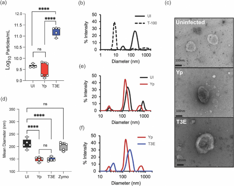
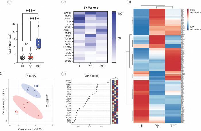
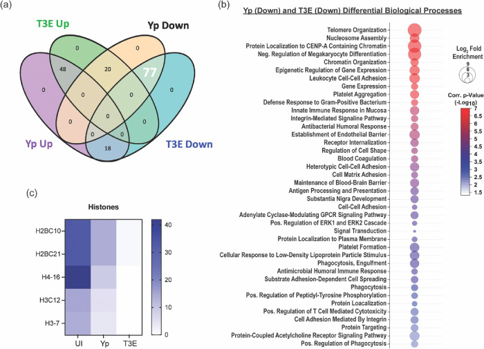
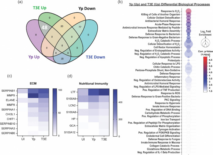
Yersinia pestis is the etiologic agent of the plague. A hallmark of plague is subversion of the host immune response by disrupting host signaling pathways required for inflammation. This non-inflammatory environment permits bacterial colonization and has been shown to be essential for disease manifestation. Previous work has shown that Y. pestis inhibits phagocytosis and degranulation by neutrophils. Manipulation of these key vesicular trafficking pathways suggests that Y. pestis influences EV secretion, cargo selection, trafficking, and/or maturation. Our goal was to define the EV population produced by neutrophils in response to Y. pestis and determine how these vesicles might influence inflammation. Toward these goals, EVs were isolated from human neutrophils infected with Y. pestis or a mutant lacking bacterial effector proteins known to manipulate host cell signaling. Mass spectrometry data revealed that cargoes packaged in EVs isolated from mutant infected cells were enriched with antimicrobials and cytotoxic proteins, contents which differed from uninfected and Y. pestis infected cells. Further, EVs produced in response to Y. pestis lacked inflammatory properties observed in those isolated from neutrophils responding to the mutant. Together, these data demonstrate that Y. pestis actively inhibits the production of antimicrobial EVs produced by neutrophils, likely contributing to immune evasion.

Keywords: Yersinia pestis; Yop effectors; human neutrophils (hPMNs); plague; type 3 secretion system (T3SS).


Conflict of interest statement

Declaration of Interest Statement All authors report no conflicts of interest.

Figures

Figure 1:
Figure 1:. Characterization of Y. pestis-elicited EVs.
hPMNs were infected with Y. pestis (Yp; red) or Y. pestis lacking the Yop effectors (T3E; blue) for 1 hour prior to EV isolation via ultracentrifugation. (a) Total EV particle quantification via NTA. Each point represents EVs from an individual human donor. (b) Representative DLS analysis of EVs from uninfected hPMNs without (UI; solid black line) or with Triton-X100 treatment (T-100; dashed line). (c) Representative TEM images of EVs isolated from uninfected, Yp, or T3E-infected hPMNs. Original magnification ~40,000x. (d) Average diameter of EVs isolated from uninfected, zymosan-treated (Zymo), Yp-infected, or T3E-infected hPMNs. Each point represents EVs from an individual human donor. (e-f) Representative DLS analysis of EVs isolated from UI, Yp, or T3E-infected hPMNs. (a,d) One-way ANOVA with Tukey; ns=not significant; ****=p≤0.0001. (b,e,f) Representative results of 5 independent donors.
Figure 2:
Figure 2:. Protein content of EVs is altered by Y. pestis.
hPMNs were infected with Y. pestis (Yp; red) or Y. pestis lacking the Yop effectors (T3E; blue) for 1 hour prior to EV isolation via ultracentrifugation. (a) Protein quantification of indicated EVs. Each point represents EVs from an individual human donor. One-way ANOVA with Tukey; ns=not significant; ****=p≤0.0001. (b) Average enrichment of recognized EV markers as measured by MS (n=5). (c) Partial Least Squares Discriminant Analysis (PLS-DA) plot depicting discriminant analysis of EVs isolated from uninfected (UI), Yp, or T3E-infected hPMNs. (d) VIP scores contributing to the variance in (c). (e) Heat map depicting protein enrichment EVs isolated from uninfected (UI), Yp, or T3E-infected hPMNs (average from 5 individual donors for each group). Proteins clustered according to Ward’s Hierarchical Agglomerative Clustering Method [54].
Figure 3:
Figure 3:. EV proteins reduced in response to Y. pestis.
(a) Venn diagram highlighting the 77 proteins (white) that were enriched in EVs from UI hPMNs (p<0.05). (b) Biological process analysis predicted by DAVID [55] using the 77 enriched proteins (pathway enrichment displayed −Log10 p>2.5). (c) Prevalence of histone proteins from EVs isolated from UI, Yp-infected, or T3E-infected EVs.
Figure 4:
Figure 4:. EV proteins enriched in response to Y. pestis.
(a) Venn diagram highlighting the 48 proteins (white) that were enriched in EVs from infected hPMNs (p<0.05) compared to the UI EV population. (b) Biological process analysis predicted by DAVID [55] using the 48 enriched proteins (pathway enrichment displayed −Log10 p>1.5). Prevalence of ECM proteins (c) and nutritional immunity proteins (d) from EVs isolated from UI, Yp-infected, or T3E-infected EVs.
Figure 5:
Figure 5:. Y. pestis T3SS limits antimicrobial and pro-inflammatory protein packaging within EVs.
(a) Venn diagram highlighting the 20 proteins (white) that were enriched in T3E EVs and subsequently reduced in Yp EVs (p<0.05) compared to the UI EV population. (b) Biological process analysis predicted by DAVID [55] using the 20 dysregulated proteins (pathway enrichment displayed −Log10 p>1.5). (c) Fold change of the 20 identified proteins relative to UI. Prevalence of Annexins (d) and antimicrobial proteins (e) from EVs isolated from UI, Yp-infected, or T3E-infected EVs.
Figure 6:
Figure 6:. Yp-elicited EVs have limited antimicrobial capacity.
(a) Y. pestis was treated for 40 min with EVs isolated from UI, Yp-infected, or T3E-infected hPMNs and bacterial viability was determined by serial dilution and conventional colony forming unit (CFU) formation on agar plates. (b) hMDMs were treated with EVs isolated from UI, Yp-infected, or T3E-infected hPMNs or IFNγ for 24 h and M1 polarization was determined by flow cytometry. (c) hMDMs were treated with EVs isolated from UI, Yp-infected, or T3E-infected hPMNs or IFNγ for 24 h and subsequently infected with Yp at an MOI of 10. Intracellular bacterial survival was assessed with conventional gentamicin protection assay followed by CFU enumeration on agar plates. One-way ANOVA with Tukey’s posttest; ns=not significant; *=p≤0.05; **= p≤0.01; ***=p≤0.001; ****=p≤0.0001
Figure 7:
Figure 7:. Yop effector proteins work cooperatively to manipulate EV protein packaging.
hPMNs were infected with WT Y. pestis (Yp) or with a Y. pestis mutant expressing only YopE (E+), YopH (H+), or YopK (K+), or mixed at a 1:1 ratio for YopE and YopH (E+H), YopH and YopK (H+K), YopE and YopK (E+K). (a) Total protein in each EV sample was quantified and normalized to the T3E control to account for donor variability. One-way ANOVA with Dunnett’s multiple comparisons test to Yp with Geisser-Greenhouse correction; **= p≤0.01; ****=p≤0.0001 (n=5–10). (b) PLS-DA plot depicting discriminant analysis of EVs from one-dimensional reverse phase liquid chromatography tandem mass spectrometry (n=5).

References

    1. Perry R.D. and Fetherston J.D., Yersinia pestis--etiologic agent of plague. Clin Microbiol Rev, 1997. 10(1): p. 35–66. - PMC - PubMed
    1. Stenseth N.C., et al. , Plague: past, present, and future. PLoS Med, 2008. 5(1): p. e3. - PMC - PubMed
    1. Inglesby T.V., et al. , Plague as a biological weapon: medical and public health management. Working Group on Civilian Biodefense. JAMA, 2000. 283(17): p. 2281–90. - PubMed
    1. Bubeck S.S., Cantwell A.M., and Dube P.H., Delayed inflammatory response to primary pneumonic plague occurs in both outbred and inbred mice. Infect Immun, 2007. 75(2): p. 697–705. - PMC - PubMed
    1. Lathem W.W., et al. , Progression of primary pneumonic plague: a mouse model of infection, pathology, and bacterial transcriptional activity. Proc Natl Acad Sci U S A, 2005. 102(49): p. 17786–91. - PMC - PubMed

Publication types

LinkOut - more resources