Review
. 2024 Dec 31:19:14171-14191.
doi: 10.2147/IJN.S493987. eCollection 2024.

Bioactive Materials Facilitate the Restoration of Neurological Function Post Cerebral Ischemic Stroke

Affiliations
Review

Bioactive Materials Facilitate the Restoration of Neurological Function Post Cerebral Ischemic Stroke

Chunyan Wang et al. Int J Nanomedicine. .

Abstract

The recovery process following ischemic stroke is a complex undertaking involving intricate cellular and molecular interactions. Cellular dysfunction or aberrant pathways can lead to complications such as brain edema, hemorrhagic transformation, and glial scar hyperplasia, hindering angiogenesis and nerve regeneration. These abnormalities may contribute to long-term disability post-stroke, imposing significant burdens on both families and society. Current clinical interventions primarily focus on endovascular therapy, overlooking the protection of brain cells themselves. However, the use of bioactive materials in stroke management has shown notable safety and efficacy. By precisely targeting the ischemic site at a cellular and molecular level, this therapeutic approach mitigates ischemia-induced brain tissue damage and promotes site repair. This review examines the protective benefits of bioactive materials in reducing cell damage and facilitating nerve restoration in accordance with the pathophysiological basis of ischemic stroke. Enhanced understanding of ischemic stroke mechanisms has the potential to advance the targeted and efficient clinical use of bioactive materials.

Keywords: angiogenesis; bioactive materials; inflammation; ischemic stroke; nerve regeneration; oxidative stress.


Conflict of interest statement

The authors report no conflicts of interest in this work.

Figures
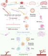
Scheme 1
Scheme 1
Treatment of ischemic stroke mediated by bioactive materials. The Scheme was created in BioRender. Wang, C. (2025) https://BioRender.com/t09f458.
Figure 1
Figure 1
Cerium oxide nanoparticles are utilized for targeted drug delivery to ischemic regions in stroke therapy. (A) The synthesis protocol for NBP-CeO2 NPs. (B) XPS analysis of Ce 3 d showed the binding energy level of Ce (III) in NBP-CeO2 NPs. (C) Quantitative representation of the proportion of BMVECs containing mitochondrial fragments (n = 3; ***P < 0.001, &P < 0.05, ##P < 0.01, ###P < 0.001). (D) Assess the ATP content in BMVECs to elucidate mitochondrial function (n = 3; ***P < 0.001, &P < 0.05, ##P < 0.01, ###P < 0.001). (E) Representative swim trajectories in the spatial learning evaluation segment of Morris water maze test. Mice locate the underwater platform as the escape latency period 22–26 days after MCAO/R (n = 12; ***P < 0.001, ###P < 0.001, &&&P < 0.001). Reprinted from Biomaterials, Li X, Han Z, Wang T, et al. Cerium oxide nanoparticles with antioxidative neurorestoration for ischemic stroke. Biomaterials. 2022;291:121904. doi:10.1016/j.biomaterials.2022.121904. Copyright 2022, with permission from Elsevier.
Figure 2
Figure 2
Polysaccharide sulfate-based nanocarriers deliver targeted neuroprotective agent rapamycin in the management of cerebral infarction. (A) The schematic design of RAPA @ tRPCS. (B) Phenotypic changes in microglia upon exposure to various nanoparticles (n = 3; *P < 0.05, ***P < 0.005). (C) The effect of different nanoparticles on microglia size (n = 3; *P < 0.05, **P < 0.01, ****P < 0.001). (D) The infarct volumes at 7 days after tMCAO were measured with ImageJ in different groups (n = 3; **P < 0.01, ***P < 0.005, ****P < 0.001). Reprinted with permission from Cao Y, Yu Y, Pan L, et al. Sulfated polysaccharide-based nanocarrier drives microenvironment-mediated cerebral neurovascular remodeling for ischemic stroke treatment. Nano Lett. 2024;24(17):5214–5223. Copyright 2024, American Chemical Society.
Figure 3
Figure 3
A APTS significantly reduces the formation of NETs by reprogramming neutrophil NETosis to cell apoptosis. (A) Preparation process of APTS. (B) The number of NETs after different treatments (n = 6; ***P < 0.001). (C) Quantitative analysis of dead cell counts in different treatments (n = 6; ***P < 0.001). (D) Schematic diagram of Morris water maze test. Path length in different processing (n = 8; ***P < 0.001). Reproduced from Yin N, Wang W, Pei F, et al. A neutrophil hijacking nanoplatform reprograming NETosis for targeted microglia polarizing mediated ischemic stroke treatment. Adv Sci. 2024;5:e2305877. http://creativecommons.org/licenses/by/4.0/. Copyright 2024, The Authors. Advanced Science published by Wiley‐VCH GmbH.
Figure 4
Figure 4
Targeting mRNA nanoparticles to improve BBB damage after ischemic stroke. (A) Schematic diagram of targeting mIL-10@MLNPs. (B) The average particle size of mIL-10@LNPs and mIL-10@MLNPs (left). Size of mIL-10@LNPs in 10% serum condition at 37 °C for up to 24 h (right). (C) Representative histograms of CD206+microglia isolated from the ischemic hemisphere of the designated treatment group. (D) Quantitative analysis of IgG leakage MFI in each group (n = 4; ****P < 0.0001). (E) Quantitative assessment of SMI32/MBP MFI ratio in the outer capsule of the indicator group (n = 4; ****P < 0.0001). Reprinted with permission Gao M, Li Y, Ho W, et al. Targeted mRNA nanoparticles ameliorate blood-brain barrier disruption postischemic stroke by modulating microglia polarization. ACS Nano. 2024;18(4):3260–3275. Copyright 2024, American Chemical Society.
Figure 5
Figure 5
SDF-1 bound nH promote angiogenesis after stroke by recruiting progenitor cells for differentiation. (A) Covalent binding of SDF-1 soluble growth factor with nH. (B) ELISA data combining SDF-1 and nH measurements and the ratio of SDF-1 to nH. (C) Quantification of NPC dissemination following 24 hours of exposure to nH, soluble SDF-1, bound and unbound SDF-1 nH at 200 ng SDF-1 (n = 3; *P < 0.05, ** P < 0.01, ***P < 0.005). (D) The perfusion vascular area in the infarcted area (left). The perfusion vascular area around the peri-infarct area (right) (n = 5; ****P < 0.001). Reproduced with permission from Wilson KL, Joseph NI, Onweller LA, et al. SDF-1 bound heparin nanoparticles recruit progenitor cells for their differentiation and promotion of angiogenesis after stroke. Adv Healthc Mater. 2023;27:e2302081. © 2023 Wiley-VCH GmbH.
Figure 6
Figure 6
The bioorthogonal MSC bioengineering system promotes neuronal regeneration after ischemic stroke through hormone effects. (A) Schematic diagram for preparing LA-HM-NP-MSC. (B) The ability of LA-HM-NP-MSC to remove ROS. (C) The PC absorption capacity of LA-HM-NP-MSC relative to natural MSC. (D) Quantitative evaluation of mitochondrial membrane potential (left) (n = 3; *P < 0.05, ***P < 0.001). Quantitative analysis of cell proliferation (right) (n = 3; *P < 0.05, ***P < 0.001). (E) Infarct size in stroke rats 28 days after MSC treatment (n = 5; ***P < 0.001, ****P < 0.0001). (F) Representative NeuN immunofluorescence images of the infarcted region following 2 months of MSC therapy (scale bar = 100 µm). Reproduced from Xu J, Sun Y, You Y, et al. Bioorthogonal microglia-inspired mesenchymal stem cell bioengineering system creates livable niches for enhancing ischemic stroke recovery via the hormesis. Acta Pharm Sin B. 2024;14(3):1412–1427. (http://creativecommons.org/licenses/by-nc-nd/4.0/). Copyright 2024, The Authors. Published by Elsevier B.V. on behalf of Chinese Pharmaceutical Association and Institute of Materia Medica, Chinese Academy of Medical Sciences.

Similar articles

Cited by

References

    1. Walter K. What is acute ischemic stroke? JAMA. 2022;327(9):885. doi:10.1001/jama.2022.1420 - DOI - PubMed
    1. Qin C, Yang S, Chu YH, et al. Signaling pathways involved in ischemic stroke: Molecular mechanisms and therapeutic interventions. Signal Transduct Target Ther. 2022;7(1):215. doi:10.1038/s41392-022-01064-1 - DOI - PMC - PubMed
    1. Singhal AB, Biller J, Elkind MS, et al. Recognition and management of stroke in young adults and adolescents. Neurology. 2013;81(12):1089–1097. doi:10.1212/WNL.0b013e3182a4a451 - DOI - PMC - PubMed
    1. Zhao Y, Hua X, Ren X, et al. Increasing burden of stroke in China: A systematic review and meta-analysis of prevalence, incidence, mortality, and case fatality. Int J Stroke. 2023;18(3):259–267. doi:10.1177/17474930221135983 - DOI - PubMed
    1. Pu L, Wang L, Zhang R, Zhao T, Jiang Y, Han L. Projected global trends in ischemic stroke incidence, deaths and disability-adjusted life years from 2020 to 2030. Stroke. 2023;54(5):1330–1339. doi:10.1161/strokeaha.122.040073 - DOI - PubMed

MeSH terms

Substances