

doi: 10.1002/ccr3.70074.
eCollection 2025 Jan.
Premature Constriction of Fetal Ductus Arteriosus Caused by Sertraline in a Pregnant Woman: A Case Report
Affiliations
- PMID: 39764265
- PMCID: PMC11702413
- DOI: 10.1002/ccr3.70074
Item in Clipboard
Premature Constriction of Fetal Ductus Arteriosus Caused by Sertraline in a Pregnant Woman: A Case Report
Clin Case Rep.
.
Abstract
Fetal ductus arteriosus was treated in a 39-year-old pregnant woman in the 33rd week After psychiatric consultation and discontinuation of sertraline which underscores the association of sertraline with premature ductus arteriosus constriction.
Keywords: case report; ductus arteriosus; pregnancy; pregnant woman; sertraline.
© 2025 The Author(s). Clinical Case Reports published by John Wiley & Sons Ltd.
Conflict of interest statement
The authors declare no conflicts of interest.
Figures
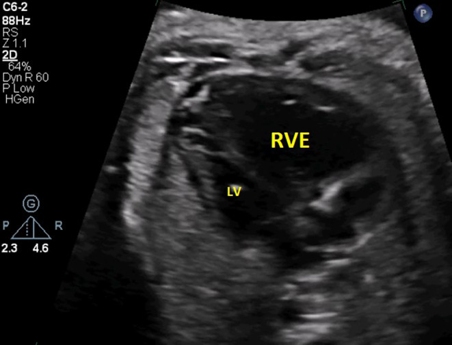
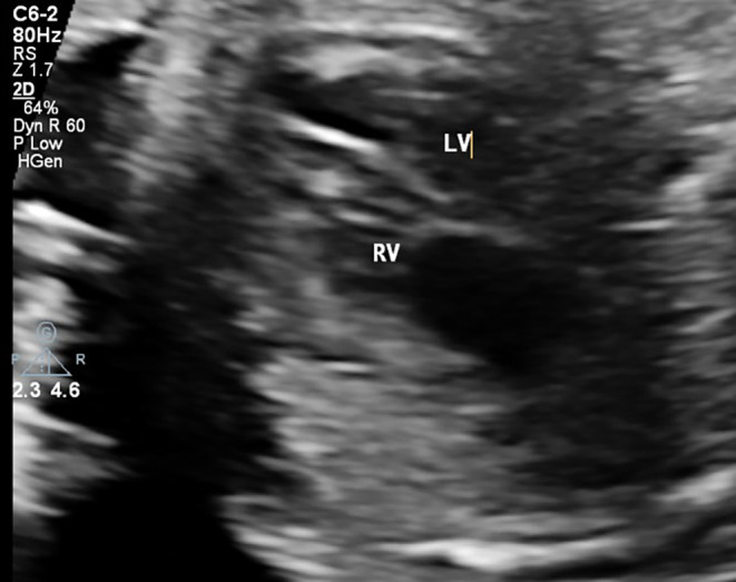
The four‐chamber view of the fetal heart demonstrates right ventricular enlargement (RVE) with a notable size discrepancy between the right ventricle (RV) and left ventricle (LV).
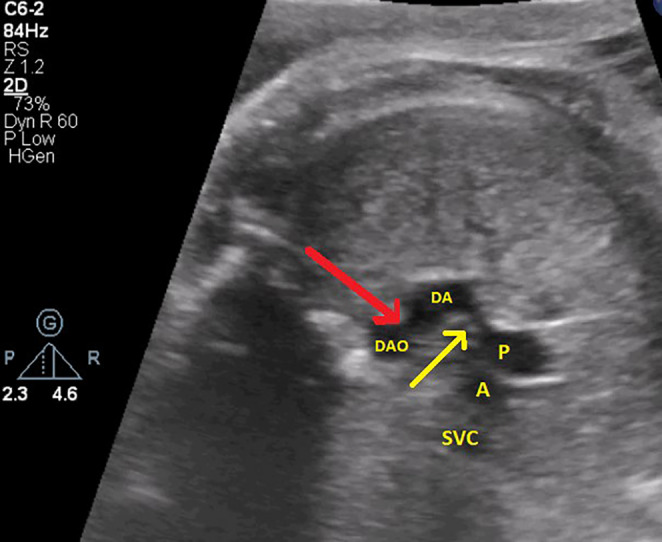
Axial imaging of the cardiac vessels reveals an S‐shaped ductus arteriosus. Notable stenosis is observed at two critical sites: the connection between the pulmonary artery (P) and the ductus arteriosus (DA) (indicated by the yellow arrow) and the junction of the ductus arteriosus with the descending aorta (DAO) (indicated by the red arrow).
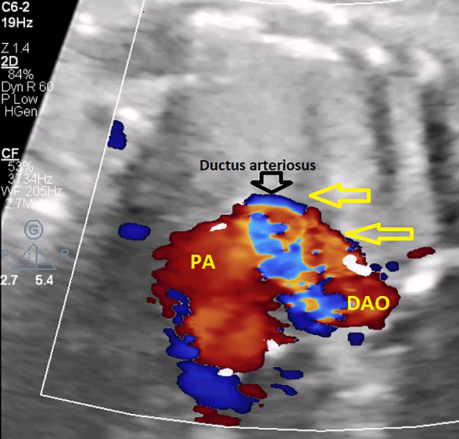
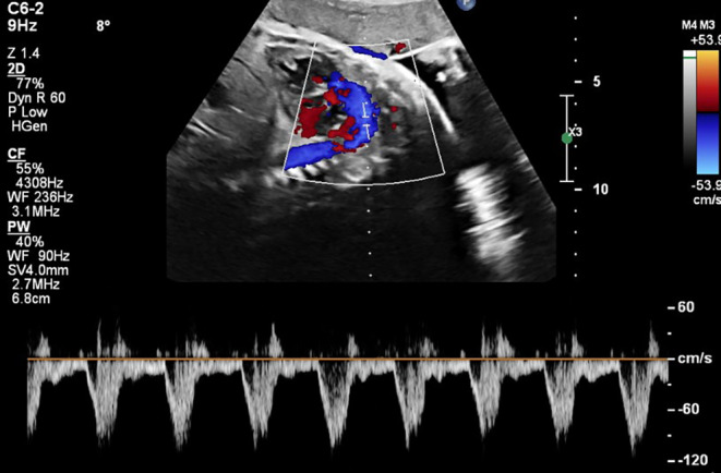
The four‐chamber view with color Doppler imaging demonstrates flow acceleration, evidenced by aliasing.
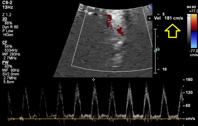
Spectral Doppler interrogation at the stenotic segment of the ductus arteriosus reveals elevated peak flow velocity, consistent with hemodynamic narrowing.
Axial fetal ultrasound demonstrates normalization of right ventricular size and resolution of the previously noted size discrepancy between the right ventricle (RV) and left ventricle (LV), with reversal of the disproportion.
Spectral Doppler evaluation of the ductus arteriosus demonstrates decreased flow velocity, indicative of a significant reduction in stenosis severity.
References
-
- Braverman A. C. and Beardslee M. A., “CHAPTER 11 ‐ The Bicuspid Aortic Valve,” In Valvular Heart Disease: A Companion to Braunwald's Heart Disease, eds. Otto C. M. and Bonow R. O. 3 ed., (Philadelphia: W.B. Saunders, 2009), 169–186.
LinkOut - more resources
Full Text Sources
