. 2024 Dec 23:15:1513311.
doi: 10.3389/fphys.2024.1513311. eCollection 2024.

Wooden breast myopathy is characterized by satellite cell dysfunction and syndecan-4 shedding

Affiliations

Wooden breast myopathy is characterized by satellite cell dysfunction and syndecan-4 shedding

Lucie Pejšková et al. Front Physiol. .

Abstract

Introduction: Skeletal muscle satellite cells (MuSCs or stem cells) play a crucial role in muscle development, maintenance, and regeneration, supporting both hypertrophy and regenerative myogenesis. Syndecans (SDCs) act as communication bridges within the muscle microenvironment, regulating interactions with extracellular matrix components and contributing significantly to tissue repair and inflammation. Specifically, syndecan-4 (SDC4) is involved in muscle regeneration at multiple stages.

Methods: This study delves into the emerging challenge of wooden breast (WB) myopathy and its connection with SDC4. Our hypothesis proposes that disruptions in MuSC dynamics through SDC4 contribute to the increased incidence of breast myopathies observed in growing broilers. To test our hypothesis, non-affected and affected broilers were systematically selected, and the characteristics of WB myopathy were studied both in vitro and in vivo. SDC4 overexpression in MuSCs and blocking peptides (BPs) corresponding to the SDC4 ectodomain were used for investigating the role of SDC4 in muscle development and its shedding levels.

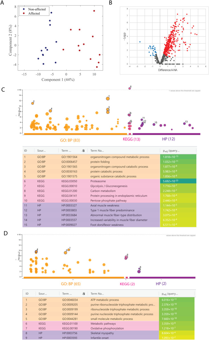
Results and discussion: In vivo examination of affected muscles revealed smaller fibers and changes in metabolic pathways. In vitro studies unveiled disrupted proliferation of MuSCs in WB myopathy, accompanied by the downregulation of several muscle markers. Investigation of the potential role of SDC4 in the pathogenesis of WB myopathy revealed a decreased tendency in SDC4 gene expression and increased shedding of its ectodomain. Moreover, we showed that SDC4 overexpression is linked to reduced proliferation in MuSCs and affected myogenesis. We detected an impaired proliferation of WB-affected MuSCs, revealing critical insights into the dysfunctional state of these cells in myopathy. Additionally, by treating MuSCs with blocking peptides derived from the SDC4 ectodomain, we identified altered proliferation. Taken together, this work contributes with valuable knowledge on the molecular mechanisms underlying WB myopathy and the role of SDC4 in this chicken myopathy.

Keywords: broiler chicken; myopathy; skeletal muscle satellite cells; syndecan-4; syndecans; wooden breast.


Conflict of interest statement

The authors declare that the research was conducted in the absence of any commercial or financial relationships that could be construed as a potential conflict of interest.

Figures

FIGURE 1
FIGURE 1
Mass spectrometry-based proteomic analysis of salt-soluble proteins. (A) Score plot from the PCA analysis of affected (red; A) and non-affected (blue; NA) samples. (B) Volcano plot of fold change (LFQ intensity) in 12 biological replicates for significantly regulated proteins. The plot is represented as a function of statistical significance (Welch’s t-test) between affected (A) and non-affected (NA) samples. X- and Y-axes show protein ratio (log2 change) in A and NA samples and p-value (−log10), respectively. Each dot represents one protein. Significantly upregulated and downregulated proteins in A samples were colored red and blue, respectively. The false discovery rate and S0 parameters were set to 0.05 and 0.1, respectively. g: GOSt Manhattan plot of upregulated (C) and downregulated (D) proteins. The x-axis shows the GO functional terms colored by data source (Gene Ontology: biological process, GO: BP; Kyoto Encyclopedia of Genes and Genomes, KEGG; Human Phenotype Ontology, HPO). Each colored dot presents a GO term. The y-axis is the adjusted −log10 p-values. The GO terms with the five lowest p-values were highlighted with a circle and listed in the table below the Manhattan plot. p-values in the table are color-coded according to significant levels, light green (less insignificant) to blue (highly significant).
FIGURE 2
FIGURE 2
Altered muscle fiber sizes in affected chicken muscle sections and PAX7. (A) Representative images of DAPI (blue)/WGA (red) counterstaining of nuclei and muscle fibers in chicken muscle sections. A marked increase in WGA staining (fibrosis) is observed in affected muscle sections. Smaller fibers can also be observed in affected muscle sections by visual inspection. Magnification: ×10 with a fluorescent microscope. P, perimysium; E, endomysium; MF, muscle fiber; FT, fibrotic tissue; N, nuclei. (B) Bars represent quantification of centrally nucleated myofibers of affected and non-affected samples images presented in (A). (C) Quantification of images in (A) show minimum Feret diameter distribution in affected and non-affected samples. (D) Representative images of PAX7/PCNA co-immunostaining in chicken muscle sections (left). Quantification of images shows that total PAX7 expressing satellite cells per 100 fibers are significantly increased in animals affected by WB; however, the fraction of proliferating PAX7+ cells are unchanged. A total of 10 random fields were quantified per image/animal. Pink arrows indicate PAX7+PCNA+, proliferating myoblasts. Yellow arrows indicate PAX7+PCNA-, non-proliferating myoblasts, and green arrows indicate proliferating cells that are not myoblasts (Pax7-PCNA+). Scale used was 100 µm. Significance was marked as ns > 0.05; *p ≤ 0.05; **p ≤ 0.01 with N = 10 affected and 11 non-affected. A; affected, NA; non-affected.
FIGURE 3
FIGURE 3
Proliferation disruption and MAPK signaling of WB MuSCs. (A) Incucyte S3 live cell proliferation assay (10x) of primary MuSCs isolated from affected and non-affected animals. Compared to affected cell lines (except one outlier SC cell line), all non-affected cell lines reached 100% confluence. (B) MuSCs treated with MEK/ERK inhibitor and (C) p38 inhibitor, (D) protein p38, ERK1/2, their phosphorylated forms, phospho (Thr180/Tyr182)-p38 and phospho (Thr202/Tyr204)-ERK1/2, and their ratios show no difference between WB affected (A) and non-affected (NA) group. Raw data for each condition (N = 3) were normalized to reference protein GAPDH and present it as a relative value to the non-affected group. Significance level was calculated by unpaired t-test with Welsh correction (ns, p > 0.5).
FIGURE 4
FIGURE 4
RNA-seq analysis of MuSCs. (A) PCA plot of top 500 variables genes between affected and non-affected chickens. A, affected; NA, non-affected (B) Volcano plot with selected genes showing the adjusted p-values and the log2 fold change (FC) values of genes. DEGs, indicated by the red dots, were identified as genes with an adjusted p-value of ≤ 0.05 and |log2 FC| between −1 and 1, and blue dots indicate only p-value. X-axis shows downregulation in the negative range of axis, and genes in the positive range are upregulated. (C) GSEA shows the top 16 pathways sorted by the normalized enrichment score, adjusted p-value (p-adjust), and gene ratio. (D) Functional enrichment analysis of the top five modules identified by network analysis.
FIGURE 5
FIGURE 5
Myogenesis and differentiation in MuSCs. (A) Gene expression of myogenesis markers obtained by RNA-seq and (B) verification of PAX7, MYOD1, and MYOG by RT-qPCR. The data are presented as the fold change average relative to the mean of non-affected WB ± SEM. Comparisons between the groups were analyzed using the t-test with Brown–Forsythe and Welsh correction; *p ≤ 0.05 (C) Differentiation assay showing intact differentiation in affected MuSCs after 2 days. Chicken NA and A MuSCs were seeded in roughly 80% confluency. Differentiated MuSCs were immunostained after 2 days with Desmin and secondary antibody Alexa Fluor™ 555 donkey anti-mouse IgG (Right panel). A, affected; NA, non-affected.
FIGURE 6
FIGURE 6
Expression and overexpression of SDC4 in chicken MuSCs. (A) Bars show the relative gene expression of SDC4 in non-affected (NA) and affected (A) chicken MuSCs (n = 8 chickens in each group) assessed by RT-qPCR. The data are presented as an average of eight chickens in triplicates. The two groups were compared using an unpaired t-test with Welch’s correction. (B) Levels of the syndecan-4 core protein (22 kDa) and two remaining syndecan-4 fragments after shedding (10 and 15 kDa) in non-affected (n = 5) and affected (n = 4) MuSCs were quantitated using ImageQuant TL and normalized to the loading control (GAPDH) and the average of NA group. Data were analyzed using an unpaired t-test with Welch’s correction and presented as a relative percentage to NA. Arrows indicate the three specific syndecan-4 bands (10, 15, and 20 kDa) confirmed by blocking experiments of the SDC4 antibody (data not shown) (ns p > 0.05; *p ≤ 0.05). (C) Co-immunostaining of SDC4 (red) and PAX7 (green) in proliferating affected and non-affected chicken MuSCs. Additionally, the panel shows staining of nuclei (blue) and overlay of the channels. (D) Gene expression levels of SDC4 in SDC4-transfected MuSCs vs. lipofectamine-treated and non-transfected/untreated MuSCs (controls). (E) Proliferation assay showing decreased proliferation of SDC4 overexpressing MuSCs. The black arrow indicates the time point of transfection (4 days of proliferation). Lipofectamine was used as a control. N = 3 individual WB non-affected chickens. (F) Gene expression levels of the myogenic markers PAX7, MYOD1, and MYOG in SDC4 overexpressing MuSCs. Untreated and lipofectamine-treated MuSCs were used as controls. Three individual animals measured in technical triplicates. Significant differences were detected using one-way ANOVA with Brown–Forsythe and Welsh correction (**p ≤ 0.01; ***p ≤ 0.001).
FIGURE 7
FIGURE 7
Effect of syndecan-4 derived blocking peptides on MuSC proliferation and shedding. (A) Schematic illustration of chicken SDC4 with its extracellular domain showing overlapping blocking peptides. Full sequence of chicken SDC4 showing exact amino acids of each blocking peptide and their overlap (underlined). The sequence also features SDC4 transmembrane domain (pink), C-terminus (light orange), and GAG attachment sites (dark orange). Illustration used partially image source from Biorender.com. (B) Effect of syndecan-4-derived blocking peptides on the proliferation of affected and non-affected MuSCs. (C) Effect of syndecan-4 derived blocking peptides on the proliferation of SDC4-overexpressing MuSCs. Black arrows indicate the time point of treatment with the respective blocking peptides. Lipofectamine was used as a control at the same final concentration. (D) Immunoblot of the 15 kDa shed SDC4 fragment in MuSCs treated with or without the respective blocking peptides. The levels were quantified using ImageQuant TL, and values were normalized to total protein LI-COR staining (loading control) and SDC4-overexpressing MuSCs without any peptide treatment (set to 1). For comparison, untreated MuSCs and the lipofectamine control are presented. Significant differences were detected using an unpaired t-test with Welch’s correction. N = 3 for individual non-affected chickens (*p ≤ 0.05).

Similar articles

Cited by

References

    1. Afratis N. A., Nikitovic D., Multhaupt H. A., Theocharis A. D., Couchman J. R., Karamanos N. K. (2017). Syndecans–key regulators of cell signaling and biological functions. Febs J. 284 (1), 27–41. 10.1111/febs.13940 - DOI - PubMed
    1. Andrews S. (2010). “FastQC: a quality control tool for high throughput sequence data,” in Babraham bioinformatics. Cambridge, United Kingdom: Babraham Institute.
    1. Andrews S. (2023). FastQC version 0.12.1. Babraham Bioinforma. Available at: https://www.bioinformatics.babraham.ac.uk/projects/fastqc/.
    1. Arnold L. L., Cecchini A., Stark D. A., Ihnat J., Craigg R. N., Carter A., et al. (2020). EphA7 promotes myogenic differentiation via cell-cell contact. Elife 9, e53689. 10.7554/eLife.53689 - DOI - PMC - PubMed
    1. Bernet J. D., Doles J. D., Hall J. K., Kelly Tanaka K., Carter T. A., Olwin B. B. (2014). p38 MAPK signaling underlies a cell-autonomous loss of stem cell self-renewal in skeletal muscle of aged mice. Nat. Med. 20 (3), 265–271. 10.1038/nm.3465 - DOI - PMC - PubMed

LinkOut - more resources