

Cell-free supernatant of Levilactobacillus brevis (RAMULAB51) from coconut inflorescence sap (Neera) enhances glucose uptake and PPAR-γ in 3T3-L1 adipocytes and inhibits α-glucosidase and α-amylase
- PMID: 39764448
- PMCID: PMC11701883
- DOI: 10.3389/fmicb.2024.1497023
Cell-free supernatant of Levilactobacillus brevis (RAMULAB51) from coconut inflorescence sap (Neera) enhances glucose uptake and PPAR-γ in 3T3-L1 adipocytes and inhibits α-glucosidase and α-amylase
Abstract
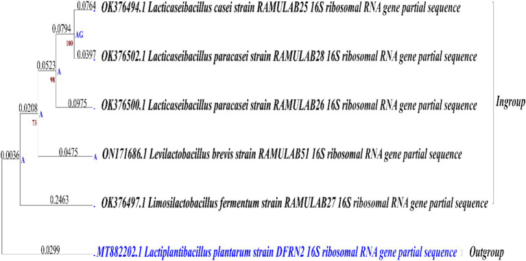
Introduction: Lactic acid bacteria are prized for their probiotic benefits and gut health improvements. This study assessed five LAB isolates from Neera, with RAMULAB51 (Levilactobacillus brevis, GenBank ON171686.1) standing out for its high hydrophobicity, auto-aggregation, antimicrobial activity, and enzyme inhibition. It evaluated RAMULAB51's potential in probiotics and diabetes management, focusing on its effects on digestive enzyme inhibition, glucose uptake, and adipocyte function.
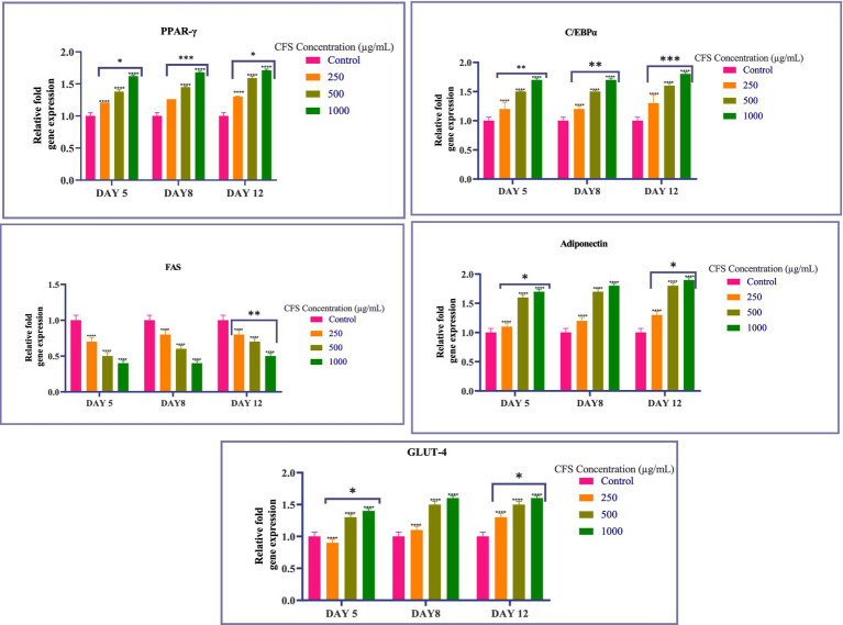
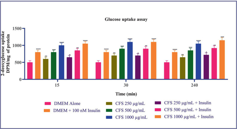
Methods: Isolates were characterized by Gram staining, catalase reaction, growth at 37°C, and tolerance to phenol, pH, and gastrointestinal conditions. Molecular identification using 16S rRNA sequencing. Evaluations included hydrophobicity, auto-aggregation, HT-29 cell line adhesion, antimicrobial activity, and antibiotic susceptibility. Enzyme inhibition was measured for α-glucosidase and α-amylase using cell-free supernatant, cell extract, and intact cells. Adipogenesis was assessed through Oil-Red O staining, gene expression analysis (PPAR-γ, C/EBPα, Adiponectin, Glut-4, FAS), and glucose uptake assays on 3T3-L1 cells.
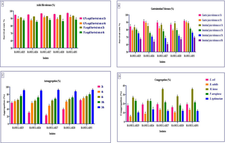
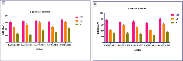
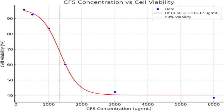
Results: All isolates showed good tolerance to pH (≤9.15 CFU/ml), phenol (≤9.90 CFU/ml), hydrophobicity (≤78.14%), and auto-aggregation (≤92.23%). RAMULAB51 demonstrated the highest tolerance, hydrophobicity, and auto-aggregation. It strongly co-aggregated with Micrococcus luteus and Bacillus subtilis, showing significant antimicrobial activity with a 24 mm inhibition zone against Micrococcus luteus. All isolates were sensitive to Ampicillin, Azithromycin, Streptomycin, and Tetracycline, but resistant to Methicillin and Vancomycin. RAMULAB51 demonstrated the highest enzyme inhibition: α-glucosidase (68.45% CFS, 60.18% CE, 42.15% IC) and α-amylase (80.74% CFS, 61.23% CE, 35.12% IC). By inhibiting these digestive enzymes, RAMULAB51 reduces the conversion of carbohydrates into glucose, thereby decreasing blood glucose levels. This reduction in circulating glucose subsequently influences adipocyte function, as evidenced by the enhanced glucose uptake (1000 µg/mL) and upregulation of PPAR-γ, C/EBPα, Adiponectin, and Glut-4, alongside the downregulation of FAS.
Conclusion: The study highlights RAMULAB51's potential for improving glucose and lipid metabolism. Further, in vivo research is needed to explore its full therapeutic benefits. These findings confirm RAMULAB51's significant probiotic potential and its promise for diabetes management, warranting further clinical investigation.
Keywords: 3T3-L1 adipocytes; Neera; PPAR-γ activation; Type 2 Diabetes Mellitus; probiotics; α-amylase; α-glucosidase.
Copyright © 2024 Kumari V B, Ramu, Shirahatti, Alam and Wong.
Conflict of interest statement
The authors declare that the research was conducted in the absence of any commercial or financial relationships that could be construed as a potential conflict of interest.
Figures
References
-
- Asha S., Asha S., Ratheesh M., Ratheesh M., Jose S. P., Jose S. P., et al. . (2019). “NEERA: a nonalcoholic nutritious beverage from unopened inflorescence of coconut palm” in Coconut-Based Nutrition and Nutraceutical Perspectives. (Singapore: Springer Nature Singapore; ) pp. 169–185.
-
- Calculator | AAT Bioquest IC50 (2023). Available at: https://www.aatbio.com/tools/ic50-calculator (Accessed June 16, 2023).
-
- Chen P., Zhang Q., Dang H., Liu X., Tian F., Zhao J., et al. . (2014). Screening for potential new probiotic based on probiotic properties and α-glucosidase inhibitory activity. Food Control 35, 65–72. doi: 10.1016/j.foodcont.2013.06.027 - DOI
LinkOut - more resources
Full Text Sources
Research Materials
Miscellaneous