. 2025 Jan 4:16:20417314241311073.
doi: 10.1177/20417314241311073. eCollection 2025 Jan-Dec.

SPI1 facilitates microfracture-mediated cartilage regeneration in the elderly by enhancing bone marrow stromal cells ctemness

Affiliations

SPI1 facilitates microfracture-mediated cartilage regeneration in the elderly by enhancing bone marrow stromal cells ctemness

Changjiang Wang et al. J Tissue Eng. .

Abstract

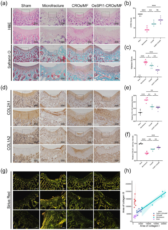
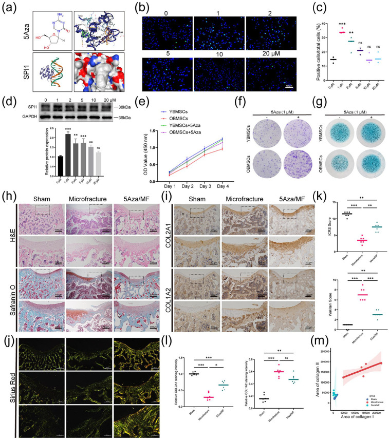
Bone marrow stimulation treatment by bone marrow stromal cells (BMSCs) released from the bone medullary cavity and differentiated into cartilage via microfracture surgery is a frequently employed technique for treating articular cartilage injuries, yet the treatment presents a main drawback of poor cartilage regeneration in the elderly. Prior research indicated that aging could decrease the stemness capacity of BMSCs, thus we made a hypothesis that increasing old BMSCs (OBMSCs) stemness might improve the results of microfracture in the elderly. First, we investigated the correlation between microfracture outcomes and BMSCs stemness using clinical data and animal experiments. The outcomes of microfracture surgery in the elderly were significantly decreased as compared with the young counterparts while the stemness capacity of OBMSCs was also significantly decreased, and they were positively correlated. To investigate the role of BMSCs stemness in microfracture, we developed microfracture-mimic cartilage regeneration organoid models. In vitro experiments identified SPI1 as a potential stemness target gene, which could enhance the stemness and chondrogenesis of OBMSCs. The implantation of cartilage regeneration organoids made by SPI1-overexpressed OBMSCs could notably enhance cartilage regeneration in the old rats as compared with the microfracture treatment alone. Furthermore, molecular docking suggested a possible interaction between SPI1 and 5-Aza-2'-deoxycytidine (5Aza). The application of 5Aza could significantly improve the result of microfracture by upregulating SPI1. In summary, we identified SPI1 as a novel stemness target of OBMSCs, which was beneficial for the improvement of microfracture-stimulated cartilage regeneration in the elderly.

Keywords: Microfracture; aging; bone marrow stromal cells; engineered organoids; stemness.


Conflict of interest statement

The author(s) declared no potential conflicts of interest with respect to the research, authorship, and/or publication of this article.

Figures

Graphical abstract
Graphical abstract
Figure 1.
Figure 1.
A significant correlation was observed between the outcomes of SD rats microfracture and the level of bone marrow stromal cells stemness. (a and b) Representative images of H&E, Safranin O/Fast green, immunohistochemical (IHC) staining of sections from rats microfracture models. n = 6. (c an d) Macroscopic scores and histological scores for assessment of the cartilage regeneration at 8 weeks after microfracture surgery. n = 6. (e and f) Quantification of COL2A1 and COL1A2 positive cells ratio in articular regenerated cartilage area. n = 6. (g) CCK-8 was performed to explore the proliferative capacity of rat YBMSCs and OBMSCs. n = 3. (h) The colony-forming abilities of YBMSCs and OBMSCs were explored by crystal violet after culture for 7 days. n = 3. (i) Spheroid formation capacity of YBMSCs and OBMSCs were explored after culture for 7 days. n = 3. (j and k) Representative immunofluorescence images and quantitative analysis of SOX9 or COL2A1 expression (green) in response to chondrogenesis treatment for 21 days. DAPI (blue) staining of nuclei. F-actin (red) staing of cytoskeleton. n = 3. (l) Strong correlation between in vitro biological properties of BMSCs, including CFU number, spheroid formation capacity and proliferative capacity, and cartilage repair measured by wakitani histology score. *p < 0.05. **p < 0.01. ***p < 0.001, ns: no significance. Values are means ± SD.
Figure 2.
Figure 2.
In comparison to YBMSCs, the expression of some specific stemness-associated genes in OBMSCs was downregulated and associated with cartilage repair. (a) Volcano plot showing the significant DEGs between human young and old groups. (b) Heat map showing the classic stemness-associated genes in two BMSCs. (c) GO analysis showing biological process, cellular compartment and molecular function of the upregulated genes involved in YBMSCs. (d) KEGG Pathway analysis showing main signaling pathways of the up-regulated genes involved in YBMSCs. (e) Venn diagram showing the overlapping genes between upregulated genes and the stemness-associated gene set. (f) qRT-PCR was performed to detect the changes in five stemness-associated molecules mRNA levels in rat BMSCs. n = 3. (g) Strong correlation between the expression levels of SPI1, WNT1, and SALL1, and wakitani histology score. *p < 0.05. **p < 0.01. ***p < 0.001, ns: no significance. Values are means ± SD.
Figure 3.
Figure 3.
Cartilage regeneration organoid models revealed that SPI1 was the most critical stemness-related gene for chondrogenesis in OBMSCs. (a) RT-qPCR was performed to verify overexpression efficiency in rat OBMSCs. n = 3. (b) Representative sections of alcian blue, SOX9, COL2A1, ACAN immunostaining. n = 3. (c) Quantification of SOX9, COL2A1, ACAN positive BMSCs in organoids. n = 3. (d and e) Representative qRT-PCR and WB of SOX9, COL2A1, and ACAN on cartilage regeneration organoids. n = 3. *p < 0.05. **p < 0.01. ***p < 0.001, ns: no significance. Values are means ± SD.
Figure 4.
Figure 4.
SPI1 regulated the stemness and chondrogenic potential of BMSCs. (a) Western blot assessed the protein expression of SPI1 on rat BMSCs after lentivirus infection. n = 3. (b) CCK-8 was performed to explore the proliferative capacity of YBMSCs, YBMSCs after knockdown of SPI1, OBMSCs, and OBMSCs after overexpression of SPI1. n = 3. (c) The colony-forming abilities of YBMSCs, YBMSCs after knockdown of SPI1, OBMSCs, and OBMSCs after overexpression of SPI1 were explored by crystal violet after culture for 7 days. n = 3. (d-e) After osteogenic induction for 21 days, ARS and ALP staining was performed in YBMSCs, YBMSCs after knockdown of SPI1, OBMSCs, and OBMSCs after overexpression of SPI1. n = 3. (f) After chondrogenic induction for 21 days, alcian blue staining was performed in YBMSCs, YBMSCs after knockdown of SPI1, OBMSCs, and OBMSCs after overexpression of SPI1. n = 3. (g and h) Representative immunofluorescence images and quantitative analysis of SOX9 (green) and COL2A1 (red) in cartilage regeneration organoids. DAPI (blue) staining of nuclei. n = 3. (i and j) Representative qRT-PCR and WB of SOX9, COL2A1, ACAN,SPI1 on four distinct types of cartilage regeneration organoids. n = 3. *p < 0.05. **p < 0.01. ***p < 0.001, ns: no significance. Values are means ± SD.
Figure 5.
Figure 5.
In vivo implantation of SPI1-overexpressed cartilage regeneration organoids could enhance cartilage repair in the old rats. (a) Representative images of H&E, Safranin O/Fast green staining of sections at 4 weeks after organoids implantation. n = 6. (b and c) Macroscopic scores and histological scores for assessment of the cartilage regeneration at 4 weeks after organoids implantation. n = 6. (d) Representative images of IHC staining of sections at 4 weeks after organoids implantation. n = 6. (e and f) Quantification of COL2A1 and COL1A2 positive cells ratio in articular regenerated cartilage area. n = 6. (g) Representative images of sirius red staining of sections at 4 weeks after organoids implantation. n = 6. (h) Quantification of type I and III collagen ratio in articular regenerated cartilage area. n = 6. *p < 0.05. **p < 0.01. ***p < 0.001, ns: no significance. Values are means ± SD.
Figure 6.
Figure 6.
5Aza could upregulate SPI1 expression and enhance cartilage regeneration in vivo and in vitro. (a) Using the RCSB model server to predict SPI1 protein structure and using CB-DOCK2 to simulate 5Aza docking with SPI1. (b and c) Representative immunofluorescence images and quantitative analysis of SPI1 in OBMSCs treated with varying concentrations of 5Aza. n = 3. (d) Representative WB images of OBMSCs treated with varying concentrations of 5Aza. n = 3. (e) CCK-8 was performed to explore the proliferative capacities of four different types of BMSCs. n = 3. (f) The colony-forming abilities of four different types of BMSCs were explored by crystal violet. n = 3. (g) Representative alcian blue staining images of four different types of BMSCs. n = 3. (h) Representative images of H&E, Safranin O/Fast green staining of sections at 4 weeks after intramuscular injection. n = 6. (i) Representative images of IHC staining of sections at 4 weeks after intramuscular injection. n = 6. (j) Representative images of sirius red staining of sections at 4 weeks after intraperitoneal injection of 5Aza. n = 6. (k) Macroscopic scores and histological scores for assessment of the cartilage regeneration at 4 weeks after intramuscular injection. n = 6. (l) Quantification of COL2A1 and COL1A2 positive cells ratio in articular regenerated cartilage area. n = 6. (m) Quantification of type I and III collagen ratio in articular regenerated cartilage area. n = 6. *p < 0.05. **p < 0.01. ***p < 0.001, ns: no significance. Values are means ± SD.

Similar articles

References

    1. Murphy MP, Koepke LS, Lopez MT, et al.. Articular cartilage regeneration by activated skeletal stem cells. Nat Med 2020; 26: 1583–1592. - PMC - PubMed
    1. Gao Y, Liu S, Huang J, et al.. The ECM-Cell Interaction of Cartilage Extracellular Matrix on Chondrocytes. BioMed Res Int 2014; 2014: 648459. - PMC - PubMed
    1. Muthu S, Korpershoek JV, Novais EJ, et al.. Failure of cartilage regeneration: emerging hypotheses and related therapeutic strategies. Nat Rev Rheumatol 2023; 19: 403–416. - PubMed
    1. Valderrabano V, Miska M, Leumann A, et al.. Reconstruction of osteochondral lesions of the talus with autologous spongiosa grafts and autologous matrix-induced chondrogenesis. Am J Sports Med 2013; 41: 519–527. - PubMed
    1. McIlwraith CW, Frisbie DD, Rodkey WG, et al.. Evaluation of intra-articular mesenchymal stem cells to augment healing of microfractured chondral defects. Arthroscopy 2011; 27: 1552–1561. - PubMed

LinkOut - more resources