Case Reports
. 2024 Dec 23:11:1522525.
doi: 10.3389/fmed.2024.1522525. eCollection 2024.

Case report: Toxic epidermal necrolysis induced by tislelizumab in a patient with esophageal squamous cell carcinoma

Affiliations
Case Reports

Case report: Toxic epidermal necrolysis induced by tislelizumab in a patient with esophageal squamous cell carcinoma

Shilong Wu et al. Front Med (Lausanne). .

Abstract

Background: Immune checkpoint inhibitors (ICIs) have been widely applicated for the treatment of patients with advanced esophageal cancer. Skin-related adverse reactions are frequent with ICIs, with toxic epidermal necrolysis (TEN) being a severe and potentially life-threatening cutaneous reaction.

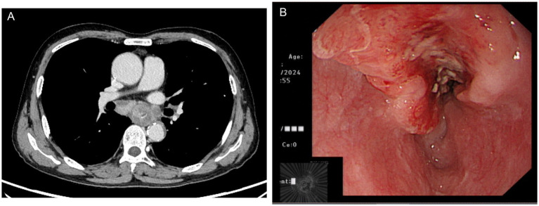
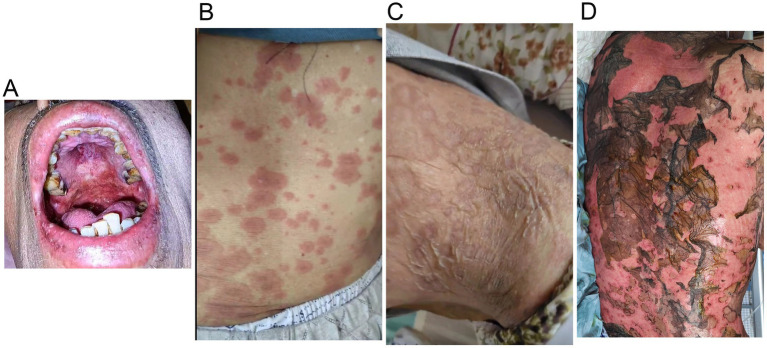
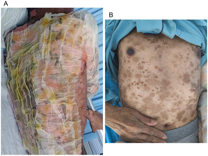
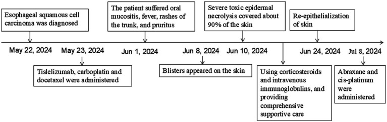
Case presentation: We present a case of a 70-year-old male with locally advanced esophageal cancer who developed severe toxic epidermal necrolysis (TEN) after 18 days of tislelizumab combined with chemotherapy. The condition rapidly progressed to cover approximately 90% of his body. After treatment with intravenous methylprednisolone, immunoglobulin, and antibiotics, along with active nutritional support and wound care, the patient recovered from TEN induced by tislelizumab.

Conclusion: Treatment for TEN is complex, and no standardized guidelines currently exist. We propose an economical, safe, effective, and simple strategy for similar TEN patients.

Keywords: esophageal squamous cell carcinoma; immune-related adverse events; skin toxicity; tislelizumab; toxic epidermal necrolysis.


Conflict of interest statement

The authors declare that the research was conducted in the absence of any commercial or financial relationships that could be construed as a potential conflict of interest.

Figures

Figure 1
Figure 1
Chest computed tomography (A) and endoscopic ultrasonography (B) showed esophageal squamous cell carcinoma in the mid-chest area.
Figure 2
Figure 2
Tislelizumab-related skin toxicity. (A) Oral mucositis; (B) Rashes of the trunk; (C) Blisters; (D) Toxic epidermal necrolysis.
Figure 3
Figure 3
Wound care for toxic epidermal necrolysis (A). An image of the skin after recovery from toxic epidermal necrolysis (B).
Figure 4
Figure 4
Schematic representation of the patient’s treatment history.

Similar articles

References

    1. Waters JK, Reznik SI. Update on Management of Squamous Cell Esophageal Cancer. Curr Oncol Rep. (2022) 24:375–85. doi: 10.1007/s11912-021-01153-4, PMID: - DOI - PubMed
    1. Shoji Y, Koyanagi K, Kanamori K, Tajima K, Ogimi M, Ninomiya Y, et al. . Immunotherapy for esophageal cancer: where are we now and where can we go. World J Gastroenterol. (2024) 30:2496–501. doi: 10.3748/wjg.v30.i19.2496, PMID: - DOI - PMC - PubMed
    1. Geisler AN, Phillips GS, Barrios DM, Wu J, Leung DYM, Moy AP, et al. . Immune checkpoint inhibitor-related dermatologic adverse events. J Am Acad Dermatol. (2020) 83:1255–68. doi: 10.1016/j.jaad.2020.03.132, PMID: - DOI - PMC - PubMed
    1. Bastuji-Garin S, Rzany B, Stern RS, Shear NH, Naldi L, Roujeau JC. Clinical classification of cases of toxic epidermal necrolysis, Stevens-Johnson syndrome, and erythema multiforme. Arch Dermatol. (1993) 129:92–6. doi: 10.1001/archderm.1993.01680220104023, PMID: - DOI - PubMed
    1. Bastuji-Garin S, Fouchard N, Bertocchi M, Roujeau JC, Revuz J, Wolkenstein P. SCORTEN: a severity-of-illness score for toxic epidermal necrolysis. J Invest Dermatol. (2000) 115:149–53. doi: 10.1046/j.1523-1747.2000.00061.x, PMID: - DOI - PubMed

Publication types

LinkOut - more resources