. 2024 Dec 12;14(24):3590.
doi: 10.3390/ani14243590.

Preliminary Findings on the Morphometric Characteristics of the Olfactory Bulb in the Cat

Affiliations

Preliminary Findings on the Morphometric Characteristics of the Olfactory Bulb in the Cat

Rui Alvites et al. Animals (Basel). .

Abstract

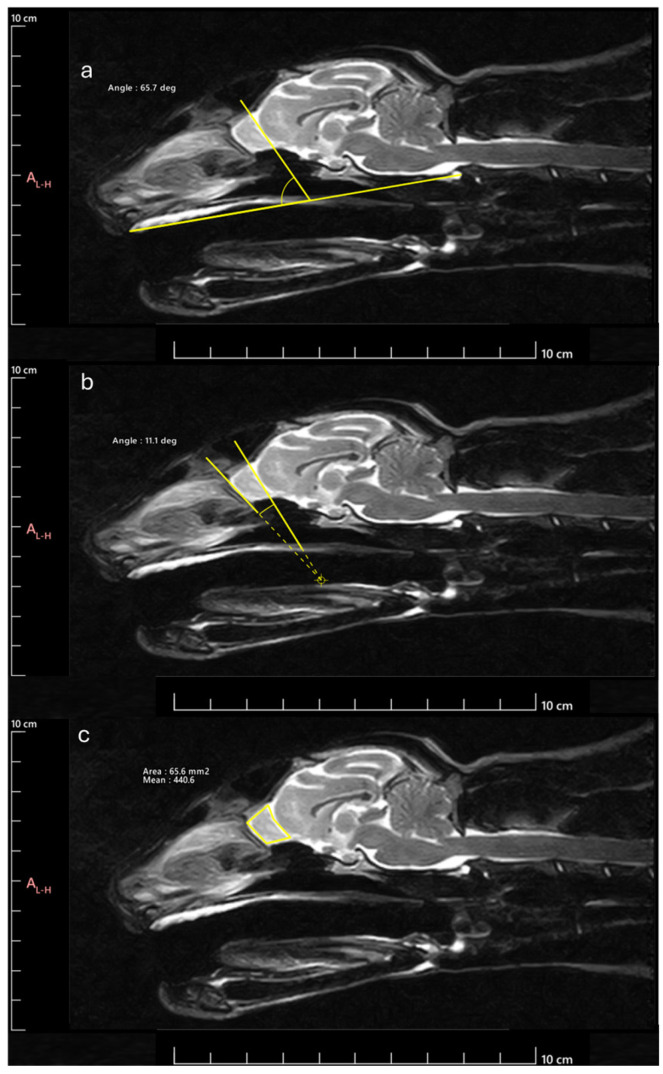
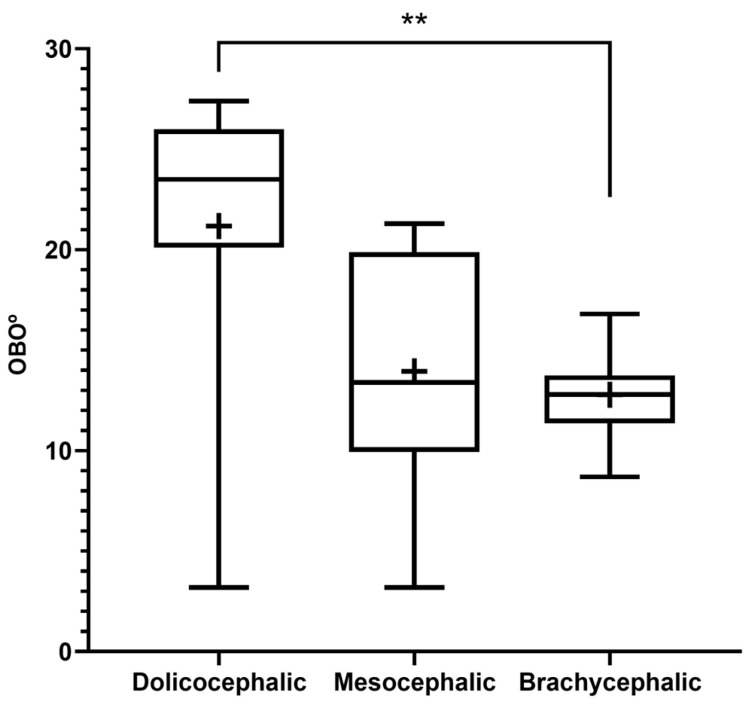
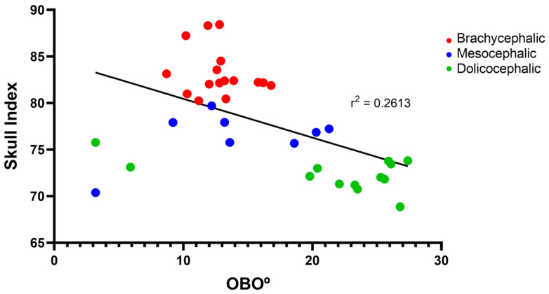
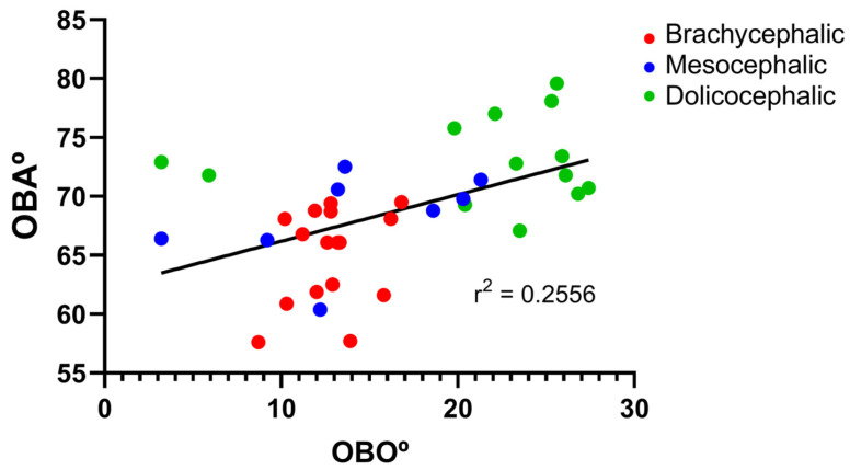
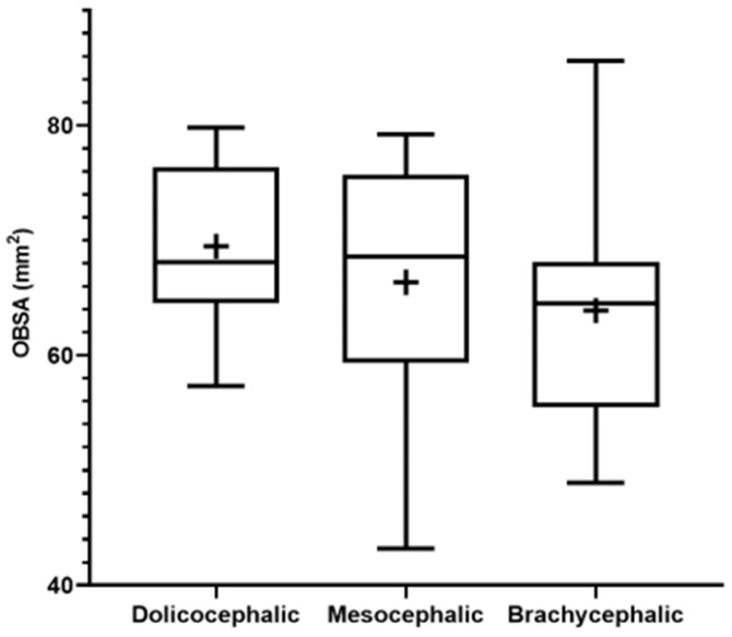
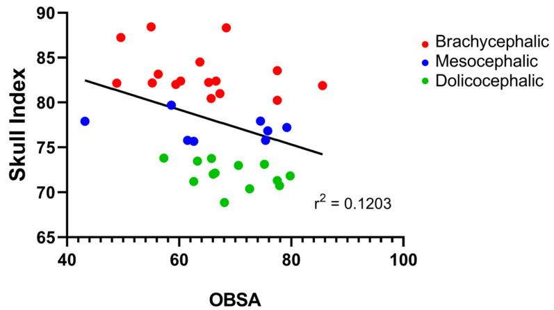
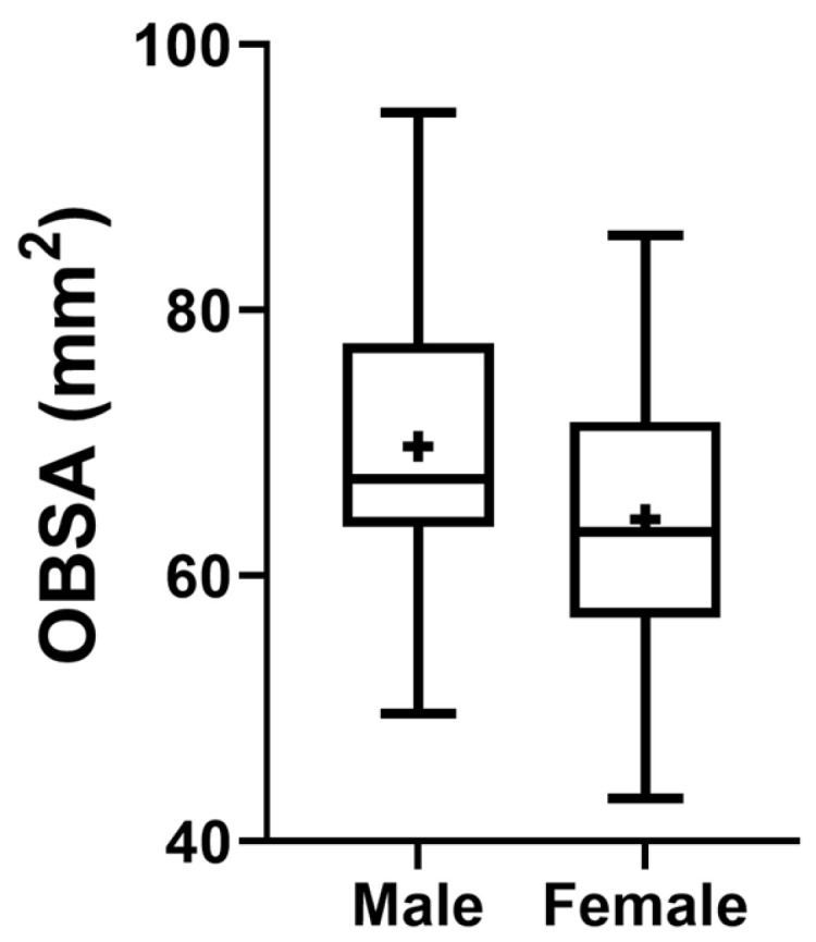
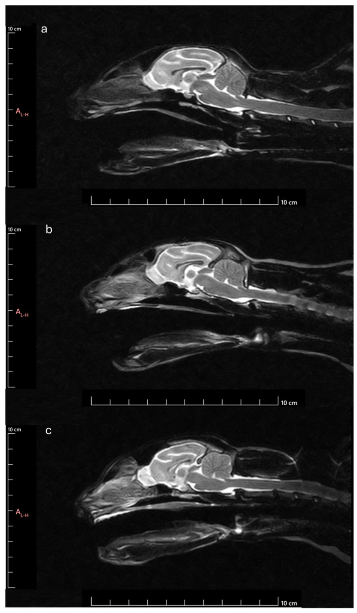
The aim of this preliminary study was to morphologically and dimensionally characterize the cat's olfactory bulb in the sagittal plane and to establish potential relationships with the cranial conformation, based on the study of in vivo MRI images. Midsagittal and transverse T2-weighted images of the head of 40 cats subjected to MRI were selected. For each animal, the skull index was calculated to classify the cranial conformation. Then, for the olfactory bulb, the angle was calculated, the orientation was determined, and the sagittal section area was measured. It was established that animals classified as brachycephalic have more compact olfactory bulbs, with smaller cross-sectional areas, ventral orientation and smaller angles established with the line that goes from the hard palate and the intercondylar notch of the foramen magnum. Animals classified as dolichocephalic have more globose and wider olfactory bulbs, dorsal orientation, and larger angles. Mesocephalic animals present an intermediate position. Males and younger adult animals have olfactory bulbs with larger cross-sectional areas than females and older animals. This work allows for the preliminarily characterization of the olfactory bulb in cats in the sagittal plane, and the correlations identified with other head structures open doors for the use of the bulb as an early indicator for the establishment of alterations of varied etiology.

Keywords: MRI; cat; companion animals; head conformation; morphometry; olfactory bulb; olfactory system.


Conflict of interest statement

The authors declare that the research was conducted in the absence of any commercial or financial relationships that could be construed as a potential conflict of interest.

Figures

Figure 1
Figure 1
Cranial measurements necessary to determine the SI: (a) SL measurement: distance between the inion and the prosthion; (b) SW measurement: distance between the outer margins of the zygomatic arches.
Figure 2
Figure 2
Methodology for determining the parameters considered: (a) OBA: angle formed by the line passing through the olfactory fissure and the line between the hard palate and the intercondylar notch of the foramen magnum; (b) OBO: open angle formed by the lines that pass through the olfactory fissure and the most rostral limit of the OB; (c) OBSA: automatically determined using the polygonal selection tool.
Figure 3
Figure 3
Box-and-whisker plots of the OBA determined from T2-weighted MRI reconstructions of the heads of 17 brachycephalic, 9 mesocephalic, and 14 dolichocephalic cats included in the study. The OBA was determined between the line passing through the olfactory fissure and the line running from the hard palate to the intercondylar notch of the foramen magnum. In each box, the middle line represents the mean value, and the lower and upper limits represent the 25th and 75th percentile, respectively. The whiskers represent the maximum and minimum values recorded. + represents the mean value. * corresponds to 0.01 ≤ p < 0.05, ** to 0.001 ≤ p < 0.01, *** to 0.0001 ≤ p < 0.001, and **** to p < 0.0001.
Figure 4
Figure 4
Scatter graphs of values of SI versus OBA determined from T2-weighted MRI reconstructions of 17 brachycephalic, 9 mesocephalic, and 14 dolichocephalic cats included in the study. The linear regression line is shown.
Figure 5
Figure 5
Box-and-whisker plots of the OBO determined from T2-weighted MRI reconstructions of the heads of 17 brachycephalic, 9 mesocephalic, and 14 dolichocephalic cats included in the study. The OBO was determined through the open angle formed by the lines that pass through the olfactory fissure and through the most rostral limit of the OB. In each box, the middle line represents the mean value, and the lower and upper limits represent the 25th and 75th percentile, respectively. The whiskers represent the maximum and minimum values recorded. + represents the mean value. * corresponds to 0.01 ≤ p < 0.05, ** to 0.001 ≤ p < 0.01, *** to 0.0001 ≤ p < 0.001, and **** to p < 0.0001.
Figure 6
Figure 6
Scatter graphs of values of SI versus OBO determined from T2-weighted MRI reconstructions of 17 brachycephalic, 9 mesocephalic, and 14 dolichocephalic cats included in the study. The linear regression line is shown.
Figure 7
Figure 7
Scatter graphs of values of OBA versus OBO determined from T2-weighted MRI reconstructions of 17 brachycephalic, 9 mesocephalic, and 14 dolichocephalic cats included in the study. The linear regression line is shown.
Figure 8
Figure 8
Box-and-whisker plots of the OBSA determined from T2-weighted MRI reconstructions of the heads of 17 brachycephalic, 9 mesocephalic, and 14 dolichocephalic cats included in the study. The OBSA was determined automatically using the polygonal selection tool of the used software. In each box, the middle line represents the mean value, and the lower and upper limits represent the 25th and 75th percentile, respectively. The whiskers represent the maximum and minimum values recorded. + represents the mean value. * corresponds to 0.01 ≤ p < 0.05, ** to 0.001 ≤ p < 0.01, *** to 0.0001 ≤ p < 0.001, and **** to p < 0.0001.
Figure 9
Figure 9
Scatter graphs of values of SI versus OBSA determined from T2-weighted MRI reconstructions of 17 brachycephalic, 9 mesocephalic, and 14 dolichocephalic cats included in the study. The linear regression line is shown.
Figure 10
Figure 10
Scatter graphs of values of OBSA versus Age determined from T2-weighted MRI reconstructions of 17 brachycephalic, 9 mesocephalic, and 14 dolichocephalic cats included in the study. The linear regression line is shown.
Figure 11
Figure 11
Box-and-whisker plots of the OBSA determined from T2-weighted MRI reconstructions of the heads of 17 brachycephalic, 9 mesocephalic, and 14 dolichocephalic cats included in the study. The OBSA was determined automatically using the polygonal selection tool of the used software. In each box, the middle line represents the mean value, and the lower and upper limits represent the 25th and 75th percentile, respectively. The whiskers represent the maximum and minimum values recorded. + represents the mean value. * corresponds to 0.01 ≤ p < 0.05, ** to 0.001 ≤ p < 0.01, *** to 0.0001 ≤ p < 0.001, and **** to p < 0.0001.
Figure 12
Figure 12
MRI Midsagittal T2-weighted images of animals with different head conformations: (a) animal classified as brachycephalic–ventral orientated and compressed OB with smaller OBSA, smaller OBA and smaller OBO; (b) Animal classified as mesocephalic-OB with intermediate orientation and OBSA, intermediate OBA and intermediate OBO; (c) Animal classified as dolichocephalic–dorsally orientated and globose OB with larger OBSA, higher OBA and higher OBO.

Similar articles

References

    1. Alvites R., Caine A., Cherubini G.B., Prada J., Varejao A.S.P., Mauricio A.C. The Olfactory Bulb in Companion Animals-Anatomy, Physiology, and Clinical Importance. Brain Sci. 2023;13:713. doi: 10.3390/brainsci13050713. - DOI - PMC - PubMed
    1. Kavoi B.M., Jameela H. Comparative morphometry of the olfactory bulb, tract and stria in the human, dog and goat. Int. J. Morphol. 2011;29:939–946. doi: 10.4067/S0717-95022011000300047. - DOI
    1. Andrews E.F., Pascalau R., Horowitz A., Lawrence G.M., Johnson P.J. Extensive Connections of the Canine Olfactory Pathway Revealed by Tractography and Dissection. J. Neurosci. 2022;42:6392–6407. doi: 10.1523/JNEUROSCI.2355-21.2022. - DOI - PMC - PubMed
    1. Hussein A. Nomenclature and Descriptive Anatomy of the Olfactory Bulb Fissure and Definition of the Olfactory Bulb Dimensions in Dogs Using in Vivo Mri. Int. J. Adv. Res. 2019;7:1120–1125. doi: 10.21474/IJAR01/9141. - DOI - PubMed
    1. Jian F., Xie D., Wu S. Characterization of Airflow Parameters in the Olfactory Fissure Zone Based on Fluid Mechanics Method. J. Craniofac. Surg. 2023;34:532–535. doi: 10.1097/SCS.0000000000009075. - DOI - PMC - PubMed

LinkOut - more resources