Case Reports
. 2024 Nov 30;13(12):997.
doi: 10.3390/biology13120997.

Fatal Feline Leukemia Virus-Associated Enteritis in a Wild Eurasian Lynx (Lynx lynx) in Germany

Affiliations
Case Reports

Fatal Feline Leukemia Virus-Associated Enteritis in a Wild Eurasian Lynx (Lynx lynx) in Germany

Katharina M Gregor et al. Biology (Basel). .

Abstract

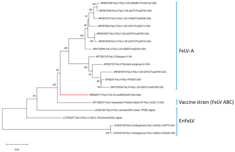
The Eurasian lynx (Lynx lynx), a widespread wild felid on the Eurasian continent, is currently classified as "critically endangered" in Germany. Understanding the impact of infectious agents is of particular importance for the continued conservation of these animals, especially regarding pathogens with broad host ranges and risk of interspecies transmission. Feline leukemia virus (FeLV) is known to infect wild and domestic felids worldwide, including several species of lynx, but it has not been reported thus far in the Eurasian lynx. In September 2020, a 16-month-old female Eurasian lynx from the Bavarian Forest, Germany, showed a sudden onset of gastrointestinal signs such as anorexia, diarrhea, and vomiting, and died within one week. Macroscopic and histologic examination revealed hemorrhagic-necrotizing enteritis and typhlocolitis, with the degeneration of crypts and crypt abscesses, as well as depleted Peyer's patches. In addition, the animal showed lymphoid depletion (lymph nodes, thymus, and spleen) and hypocellularity of the bone marrow. FeLV infection was confirmed by immunohistochemistry and next generation sequencing. A secondary bacterial infection with hemolytic Escherichia coli and Clostridium perfringens type A was present in the intestine. This is the first report of FeLV-associated enteritis, lymphoid depletion and bone marrow suppression with associated secondary bacterial infection in a Eurasian lynx.

Keywords: Eurasian lynx; FeLV-associated enteritis; feline leukemia virus; secondary bacterial infection; wildlife.


Conflict of interest statement

The authors declare no conflicts of interest. The funders had no role in the design of the study; in the collection, analyses, or interpretation of data; in the writing of the manuscript; or in the decision to publish the results.

Figures

Figure 1
Figure 1
Macroscopic findings in a Eurasian lynx (Lynx lynx). The jejunum is diffusely red and filled with blood-tinged digesta.
Figure 2
Figure 2
Histologic and immunohistochemical findings in small intestine and mesenteric lymph node of a Eurasian lynx (Lynx lynx). (a) Severe, diffuse, subacute, hemorrhagic-necrotizing enteritis with loss of intestinal crypts, crypt dilatation (star), and crypt epithelial degeneration. Hematoxylin–eosin (HE). Bar = 100 µm. Insert. Dilatation of a single intestinal crypt with crypt epithelial degeneration (arrows) and luminal accumulation of cellular debris (crypt abscess). HE. Bar = 20 µm. (b) Lymphatic depletion and blood resorption in mesenteric lymph node. HE. Bar = 100 µm. (c) Immunolabeling of feline leukemia virus gp85/gp70 envelope protein in the intestinal crypt epithelium (arrows) and infiltrating mononuclear cells (arrowheads). Bars = 50 µm. (d) Immunolabeling of feline leukemia virus gp85/gp70 envelope protein in mononuclear cells (arrowheads) in mesenteric lymph node. Bar = 50 µm.
Figure 3
Figure 3
Phylogenetic analysis based on the complete coding sequence of feline leukemia virus (FeLV). The tree was constructed using the maximum likelihood method and the GTR + G was used as best fit model to shape evolutionary rates across sites with 1000 bootstraps. Bootstrap values are presented at nodes. The scale bar indicates the number of nucleotide changes per site. Taxon names are presented by the GenBank accession number, virus name and strain. Genome sequences belonging to different FeLV clades were downloaded from GenBank (Table S4).

References

    1. Hartmann K., Hofmann-Lehmann R. What’s new in feline leukemia virus infection. Vet. Clin. N. Am. Small Anim. Pract. 2020;50:1013–1036. doi: 10.1016/j.cvsm.2020.05.006. - DOI - PubMed
    1. Hofmann-Lehmann R., Hartmann K. Feline leukaemia virus infection: A practical approach to diagnosis. J. Feline Med. Surg. 2020;22:831–846. doi: 10.1177/1098612X20941785. - DOI - PMC - PubMed
    1. Gleich S., Hartmann K. Hematology and serum biochemistry of feline immunodeficiency virus-infected and feline leukemia virus-infected cats. J. Vet. Intern. Med. 2009;23:552–558. doi: 10.1111/j.1939-1676.2009.0303.x. - DOI - PubMed
    1. Chiu E.S., Kraberger S., Cunningham M., Cusack L., Roelke M., Vande Woude S. Multiple introductions of domestic cat feline leukemia virus in endangered Florida panthers. Emerg. Infect. Dis. 2019;25:92–101. doi: 10.3201/eid2501.181347. - DOI - PMC - PubMed
    1. Mora M., Napolitano C., Ortega R., Poulin E., Pizarro-Lucero J. Feline immunodeficiency virus and feline leukemia virus infection in free-ranging guignas (Leopardus guigna) and sympatric domestic cats in human perturbed landscapes on Chiloé Island, Chile. J. Wildl. Dis. 2015;51:199–208. doi: 10.7589/2014-04-114. - DOI - PubMed

Publication types

LinkOut - more resources